 Explore
 Validate
 Learn
LearnAM06286SU-N
antibody from OriGene
Targeting: CD44
CD44R, CSPG8, HCELL, IN, MC56, MDU2, MDU3, MIC4, Pgp1
 Western blot
 ELISA
ELISAAntibody data
- Antibody Data
- Antigen structure
- References [0]
- Comments [0]
- Validations
- Western blot [2]
- Immunocytochemistry [2]
- Immunohistochemistry [2]
- Flow cytometry [1]
Submit
Validation data
Reference
Comment
Report error
- Product number
- AM06286SU-N - Provider product page

- Provider
- OriGene
- Product name
- CD44 mouse monoclonal antibody, clone 8E2F3, Ascites
- Antibody type
- Monoclonal
- Description
- CD44 mouse monoclonal antibody, clone 8E2F3, Ascites
- Host
- Mouse
- Conjugate
- Unconjugated
- Epitope
- CD44
- Isotype
- IgG
- Antibody clone number
- 8E2F3
- Vial size
- 100 µl
No comments: Submit comment
Supportive validation
- Submitted by
- OriGene (provider)
- Main image

- Experimental details
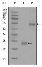
- Western blot analysis using CD44 mouse mAb against truncated Trx-CD44 recombinant protein (1) and GST-CD44 (aa628-699) recombinant protein (2).
- Validation comment
- WB
- Submitted by
- OriGene (provider)
- Main image

- Experimental details
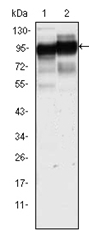
- Western blot analysis using CD44 mouse mAb against Hela (1) and HUVE-12(2) cell lysate.
- Validation comment
- WB
Supportive validation
- Submitted by
- OriGene (provider)
- Main image
- Experimental details
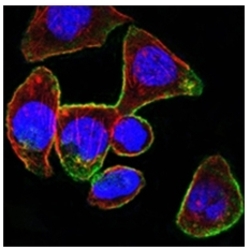
- Confocal immunofluorescence analysis of PANC-1 cells using CD44 mouse mAb (green). Red: Actin filaments have been labeled with DY-554 phalloidin. Blue: DRAQ5 fluorescent DNA dye.
- Validation comment
- IF

- Submitted by
- OriGene (provider)
- Main image

- Experimental details
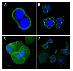
- Confocal immunofluorescence analysis of methanol-fixed A431 (A), Hela (B), PANC-1 (C) and EC (D) cells using CD44 mouse mAb (green), showing membrane localization. Blue: DRAQ5 fluorescent DNA dye.
- Validation comment
- IF
Supportive validation
- Submitted by
- OriGene (provider)
- Main image
- Experimental details
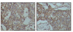
- Immunohistochemical analysis of paraffin-embedded human breast carcinoma tissues, showing membrane localization with DAB staining using CD44 mouse mAb
- Validation comment
- IHC

- Submitted by
- OriGene (provider)
- Main image

- Experimental details
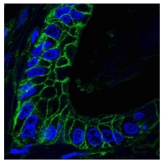
- Confocal analysis of paraffin-embedded human lung cancer tissues using CD44 mouse mAb (green), showing membrane localization. Blue: DRAQ5 fluorescent DNA dye.
- Validation comment
- IHC
Supportive validation
- Submitted by
- OriGene (provider)
- Main image
- Experimental details
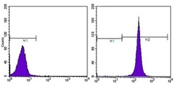
- Flow cytometric analysis of Hela cells using CD44 mouse mAb (right) and negative control (left).
- Validation comment
- FC
