Explore
 Validate
 Learn
Learn Western blot
Antibody data
- Antibody Data
- Antigen structure
- References [0]
- Comments [0]
- Validations
- Western blot [1]
- Immunohistochemistry [3]
Submit
Validation data
Reference
Comment
Report error
- Product number
- PA5-18677 - Provider product page

- Provider
- Invitrogen Antibodies
- Product name
- Gpx3 Polyclonal Antibody
- Antibody type
- Polyclonal
- Antigen
- Synthetic peptide
- Description
- This antibody is predicted to react with canine, human and rat based on sequence homology. This antibody is tested in Peptide ELISA: antibody detection limit dilution 8,000.
- Reactivity
- Human, Mouse
- Host
- Goat
- Isotype
- IgG
- Vial size
- 100 µg
- Concentration
- 0.5 mg/mL
- Storage
- -20° C, Avoid Freeze/Thaw Cycles
No comments: Submit comment
Supportive validation
- Submitted by
- Invitrogen Antibodies (provider)
- Main image
- Experimental details
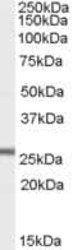
- Western Blot staining of Mouse Heart lysate using Product # PA5-18677 at a concentration of 1.0 µg/mL, the primary antibody incubation was 1 hour and the detection method was chemiluminescence.
Supportive validation
- Submitted by
- Invitrogen Antibodies (provider)
- Main image
- Experimental details
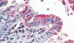
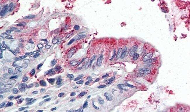
- Immunohistochemical analysis of Gpx3 in Human Colon using a Gpx3 monoclonal antibody (Product #PA5-18677) at 5 µg/mL. The Human Colon tissue section was paraffin embeded and detected using steamed antigen retrieval with citrate buffer pH 6, AP-staining.
- Submitted by
- Invitrogen Antibodies (provider)
- Main image
- Experimental details
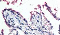
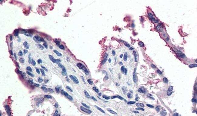
- Immunohistochemical analysis of Gpx3 in Human Placenta using a Gpx3 monoclonal antibody (Product #PA5-18677) at 5 µg/mL. The Human Placenta tissue section was paraffin embeded and detected using steamed antigen retrieval with citrate buffer pH 6, AP-staining.
- Submitted by
- Invitrogen Antibodies (provider)
- Main image
- Experimental details
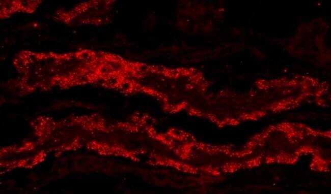
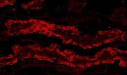
- Immunohistochemical analysis of Gpx3 in Porcine Kidney using a Gpx3 monoclonal antibody (Product #PA5-18677) at 20 µg/mL. The Porcine Kidney tissue section was PSA perfused cryosection and detected using Microwave antigen retrieval with citrate buffer pH 3, CY3-staining.