Antibody data
- Antibody Data
- Antigen structure
- References [9]
- Comments [0]
- Validations
- Western blot [6]
- Immunohistochemistry [4]
Submit
Validation data
Reference
Comment
Report error
- Product number
- NBP1-81734 - Provider product page

- Provider
- Novus Biologicals
- Proper citation
- Novus Cat#NBP1-81734, RRID:AB_11002818
- Product name
- Rabbit Polyclonal LONP1 Antibody
- Antibody type
- Polyclonal
- Description
- Immunogen affinity purified. Specificity of human LONP1 antibody verified on a Protein Array containing target protein plus 383 other non-specific proteins.
- Reactivity
- Human, Mouse, Rat, Drosophila
- Host
- Rabbit
- Isotype
- IgG
- Vial size
- 0.1 ml
- Storage
- Store at 4C short term. Aliquot and store at -20C long term. Avoid freeze-thaw cycles.
Submitted references Concentration of mitochondrial DNA mutations by cytoplasmic transfer from platelets to cultured mouse cells.
Reciprocal Roles of Tom7 and OMA1 during Mitochondrial Import and Activation of PINK1.
Lon protease inactivation in Drosophila causes unfolded protein stress and inhibition of mitochondrial translation.
Defects in the mitochondrial-tRNA modification enzymes MTO1 and GTPBP3 promote different metabolic reprogramming through a HIF-PPARγ-UCP2-AMPK axis.
Loss of the Drosophila m-AAA mitochondrial protease paraplegin results in mitochondrial dysfunction, shortened lifespan, and neuronal and muscular degeneration.
microRNA-mediated differential expression of TRMU, GTPBP3 and MTO1 in cell models of mitochondrial-DNA diseases.
PINK1-Parkin pathway activity is regulated by degradation of PINK1 in the mitochondrial matrix.
The accumulation of misfolded proteins in the mitochondrial matrix is sensed by PINK1 to induce PARK2/Parkin-mediated mitophagy of polarized mitochondria.
Proteomics analysis reveals novel components in the detergent-insoluble subproteome in Alzheimer's disease.
Ishikawa K, Kobayashi K, Yamada A, Umehara M, Oka T, Nakada K
PloS one 2019;14(3):e0213283
PloS one 2019;14(3):e0213283
Reciprocal Roles of Tom7 and OMA1 during Mitochondrial Import and Activation of PINK1.
Sekine S, Wang C, Sideris DP, Bunker E, Zhang Z, Youle RJ
Molecular cell 2019 Mar 7;73(5):1028-1043.e5
Molecular cell 2019 Mar 7;73(5):1028-1043.e5
Lon protease inactivation in Drosophila causes unfolded protein stress and inhibition of mitochondrial translation.
Pareek G, Thomas RE, Vincow ES, Morris DR, Pallanck LJ
Cell death discovery 2018;4:51
Cell death discovery 2018;4:51
Defects in the mitochondrial-tRNA modification enzymes MTO1 and GTPBP3 promote different metabolic reprogramming through a HIF-PPARγ-UCP2-AMPK axis.
Boutoual R, Meseguer S, Villarroya M, Martín-Hernández E, Errami M, Martín MA, Casado M, Armengod ME
Scientific reports 2018 Jan 18;8(1):1163
Scientific reports 2018 Jan 18;8(1):1163
Loss of the Drosophila m-AAA mitochondrial protease paraplegin results in mitochondrial dysfunction, shortened lifespan, and neuronal and muscular degeneration.
Pareek G, Thomas RE, Pallanck LJ
Cell death & disease 2018 Feb 21;9(3):304
Cell death & disease 2018 Feb 21;9(3):304
microRNA-mediated differential expression of TRMU, GTPBP3 and MTO1 in cell models of mitochondrial-DNA diseases.
Meseguer S, Boix O, Navarro-González C, Villarroya M, Boutoual R, Emperador S, García-Arumí E, Montoya J, Armengod ME
Scientific reports 2017 Jul 24;7(1):6209
Scientific reports 2017 Jul 24;7(1):6209
PINK1-Parkin pathway activity is regulated by degradation of PINK1 in the mitochondrial matrix.
Thomas RE, Andrews LA, Burman JL, Lin WY, Pallanck LJ
PLoS genetics 2014;10(5):e1004279
PLoS genetics 2014;10(5):e1004279
The accumulation of misfolded proteins in the mitochondrial matrix is sensed by PINK1 to induce PARK2/Parkin-mediated mitophagy of polarized mitochondria.
Jin SM, Youle RJ
Autophagy 2013 Nov 1;9(11):1750-7
Autophagy 2013 Nov 1;9(11):1750-7
Proteomics analysis reveals novel components in the detergent-insoluble subproteome in Alzheimer's disease.
Gozal YM, Duong DM, Gearing M, Cheng D, Hanfelt JJ, Funderburk C, Peng J, Lah JJ, Levey AI
Journal of proteome research 2009 Nov;8(11):5069-79
Journal of proteome research 2009 Nov;8(11):5069-79
No comments: Submit comment
Supportive validation
- Submitted by
- Novus Biologicals (provider)
- Main image

- Experimental details
- Simple Western: LONP1 Antibody [NBP1-81734] - Simple Western lane view shows a specific band for LONP1 in 0.2 mg/ml of h. Kidney (left) , NIH-3T3 (right) lysate. This experiment was performed under reducing conditions using the 12-230 kDa separation system.
- Submitted by
- Novus Biologicals (provider)
- Main image

- Experimental details
- Simple Western: LONP1 Antibody [NBP1-81734] - Electropherogram image(s) of corresponding Simple Western lane view. LONP1 antibody was used at 1:20 dilution on h. Kidney and NIH-3T3 lysate(s).
- Submitted by
- Novus Biologicals (provider)
- Main image

- Experimental details
- Western Blot: LONP1 Antibody [NBP1-81734] - Analysis in mouse cell line NIH-3T3, rat cell line NBT-II and rat cell line pC12.
- Submitted by
- Novus Biologicals (provider)
- Main image

- Experimental details
- Western Blot: LONP1 Antibody [NBP1-81734] - Analysis in A-431 cells transfected with control siRNA, target specific siRNA probe #1 and #2, using Anti-LONP1 antibody. Remaining relative intensity is presented. Loading control: Anti-GAPDH.
- Submitted by
- Novus Biologicals (provider)
- Main image

- Experimental details
- Western Blot: LONP1 Antibody [NBP1-81734] - Analysis using Anti-LONP1 antibody NBP1-81734 (A) shows similar pattern to independent antibody NBP2-76504 (B).
- Submitted by
- Novus Biologicals (provider)
- Main image

- Experimental details
- Western Blot: LONP1 Antibody [NBP1-81734] - PINK1 accumulates upon knockdown of Lon. Western blot analysis of Lon protease (Lon), Mitochondrial transcription factor A (TFAM), Heat shock protein 60 (Hsp60) and Actin in whole head homogenate from control and Lon deficient animals. Image collected and cropped by CiteAb from the following publication (http://dx.plos.org/10.1371/journal.pgen.1004279), licensed under a CC-BY licence.
Supportive validation
- Submitted by
- Novus Biologicals (provider)
- Main image

- Experimental details
- Immunohistochemistry-Paraffin: LONP1 Antibody [NBP1-81734] - Staining of human duodenum shows granular cytoplasmic positivity in glandular cells.
- Submitted by
- Novus Biologicals (provider)
- Main image

- Experimental details
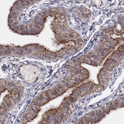
- Immunohistochemistry-Paraffin: LONP1 Antibody [NBP1-81734] - Staining of human fallopian tube shows granular cytoplasmic positivity in glandular cells.
- Submitted by
- Novus Biologicals (provider)
- Main image

- Experimental details
- Immunohistochemistry-Paraffin: LONP1 Antibody [NBP1-81734] - Staining of human pancreas shows granular cytoplasmic positivity.
- Submitted by
- Novus Biologicals (provider)
- Main image

- Experimental details
- Immunohistochemistry-Paraffin: LONP1 Antibody [NBP1-81734] - Staining of human tonsil shows granular cytoplasmic positivity.
 Explore
 Validate
 Learn
Learn Western blot
 Immunocytochemistry