Explore
 Validate
 Learn
Learn Western blot
 ELISA
ELISAAntibody data
- Antibody Data
- Antigen structure
- References [0]
- Comments [0]
- Validations
- Western blot [4]
Submit
Validation data
Reference
Comment
Report error
- Product number
- GTX48743 - Provider product page

- Provider
- GeneTex
- Proper citation
- GeneTex Cat#GTX48743, RRID:AB_11171245
- Product name
- Ajuba antibody
- Antibody type
- Polyclonal
- Reactivity
- Human
- Host
- Rabbit
No comments: Submit comment
Supportive validation
- Submitted by
- GeneTex (provider)
- Main image
- Experimental details
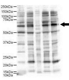
- Western blot using GeneTex's Affinity Purified anti-Ajuba antibody shows detection of a 57-kDa band consistent with the expected MW for Ajuba (arrowhead). Lanes correspond to 1) HeLa nuclear extract, and 2) HeLa, 3) A431, 4) Jurkat and 5) 293 whole cell lysates. Immunoprecipitation of Ajuba followed by western blotting may result in cleaner background staining. Approximately 5 µg of each preparation was run on a SDS-PAGE and transferred onto nitrocellulose followed by reaction with a 1:500 dilution of anti-Ajuba antibody. Detection occurred using a 1:5,000 dilution of HRP-labeled Donkey anti-Rabbit IgG for 1 hour at room temperature. A chemiluminescence system was used for signal detection (Roche) using a 60-sec exposure time. Personal Communication. E. Pugacheva, Fox Chase Cancer Center, Philadelphia, PA.
- Validation comment
- WB

- Submitted by
- GeneTex (provider)
- Main image

- Experimental details
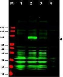
- Western blot using GeneTex's Affinity Purified anti-GGA1 antibody shows detection of bands at ~100 kDa corresponding to YFP-GGA1 fusion present in a lysate of HEK293 cells over- expressing the recombinant protein (arrowhead). Approximately 35 µg of lysate was separated on a 4-20% gel by SDS-PAGE and transferred onto nitrocellulose. After blocking the membrane was probed with the primary antibody diluted to 1:1,350. Reaction occurred overnight at 4° followed by washes and reaction with a 1:10,000 dilution of IRDye800 conjugated rabbit anti-Goat IgG [H&L] MXHu for 45 min at room temperature. IRDye800 fluorescence image was captured using the Odyssey® Infrared Imaging System developed by LI-COR. IRDye is a trademark of LI-COR, Inc. Other detection systems will yield similar results..
- Validation comment
- WB
- Submitted by
- GeneTex (provider)
- Main image
- Experimental details
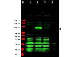
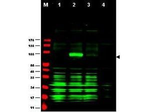
- Western blot using Affinity Purified anti-Ajuba antibody shows detection of Ajuba-RFP fusion protein in cell lysates (arrow-head). Lanes correspond to 1) vector only trans-fection, 2) human Ajuba-RFP, 3) mouse Ajuba-RFP, and 4) mock transfection. Approximately 50 ?g of each lysate was loaded per lane for SDS-PAGE followed by transfer onto nitrocellulose and reaction with a 1:1,700 dilution of anti-Ajuba antibody.
- Submitted by
- GeneTex (provider)
- Main image

- Experimental details
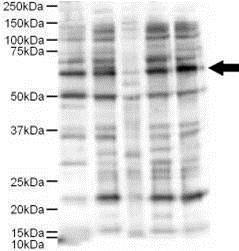
- Western blot using Affinity Purified anti-Ajuba antibody shows detection of a 57-kDa band consistent with the expected MW for Ajuba (arrowhead). Lanes correspond to 1) HeLa nuclear extract, and 2) HeLa, 3) A431, 4) Jurkat and 5) 293 whole cell lysates. Immunoprecipitation of Ajuba followed by western blotting may result in cleaner background staining. Approximately 5 ?g of each preparation was run on a SDS-PAGE and transferred onto nitrocellulose followed by reaction with a 1:500 dilution of anti-Ajuba antibody. Detection occurred using a 1:5,000 dilution of HRP-labeled Donkey anti-Rabbit IgG for 1 hour at room temperature. A chemiluminescence system was used for signal detection (Roche) using a 60-sec exposure time.