 Explore
 Validate
 Learn
Learn701830
antibody from Invitrogen Antibodies
Targeting: FERMT1
C20orf42, FLJ20116, KIND1, UNC112A, URP1
 Western blot
Antibody data
- Antibody Data
- Antigen structure
- References [0]
- Comments [0]
- Validations
- Western blot [1]
- Immunocytochemistry [1]
- Flow cytometry [1]
Submit
Validation data
Reference
Comment
Report error
- Product number
- 701830 - Provider product page

- Provider
- Invitrogen Antibodies
- Product name
- Kindlin Recombinant Rabbit Monoclonal Antibody (7H7L9)
- Antibody type
- Monoclonal
- Antigen
- Other
- Description
- This antibody is predicted to react with Monkey, Equine, Bovine, Pig and Rabbit.
- Antibody clone number
- 7H7L9
- Concentration
- 0.5 mg/mL
No comments: Submit comment
Supportive validation
- Submitted by
- Invitrogen Antibodies (provider)
- Main image

- Experimental details
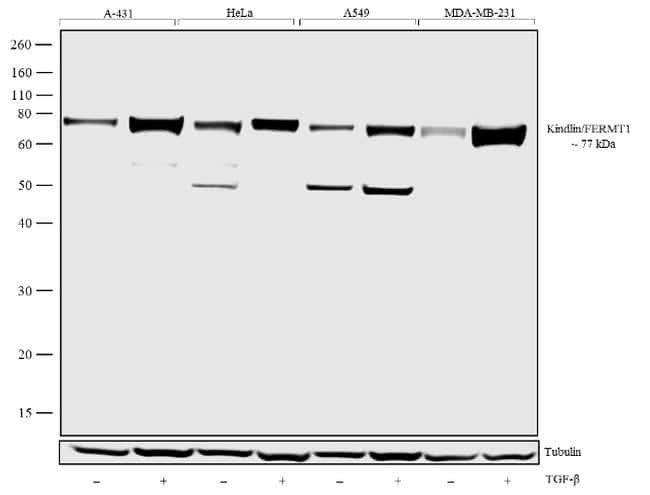
- Western blot analysis was performed on whole cell extracts (30 µg lysate) of A-431 (Lane 1), A-431 treated with TGF-beta (20 ng/mL for 15 min) (Lane 2), HeLa (Lane 3), HeLa Treated with TGF-beta (20 ng/mL for 15 min) (Lane 4), A549 (Lane 5), A549 Treated with TGF-beta (20 ng/mL for 15 min) (Lane 6), MDA-MB-231 (Lane 7), MDA-MB-231 Treated with TGF-beta (20 ng/mL for 15 min) (Lane 8). The blots were probed with Anti-Kindlin/FERMT1 Recombinant Rabbit Monoclonal Antibody (Product # 701830, 1-2 µg/mL) and detected by chemiluminescence using Goat anti-Rabbit IgG (H+L) Superclonal™ Secondary Antibody, HRP conjugate (Product # A27036, 0.4 µg/mL, 1:2500 dilution). A 77 kDa and 53 kDa bands corresponding to Kindlin/FERMT1 was observed according to the treatment. Known quantity of protein samples were electrophoresed using Novex® NuPAGE® 10% Bis-Tris gel (Product # NP0301BOX), XCell SureLock™ Electrophoresis System (Product # EI0002) and Novex® Sharp Pre-Stained Protein Standard (Product # LC5800). Resolved proteins were then transferred onto a nitrocellulose membrane with iBlot® Dry Blotting System (Product # IB21001). The membrane was probed with the relevant primary and secondary Antibody following blocking with 5% skimmed milk. Chemiluminescent detection was performed using Pierce™ ECL Western blotting Substrate (Product # 32106).
Supportive validation
- Submitted by
- Invitrogen Antibodies (provider)
- Main image
- Experimental details
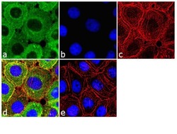
- Immunofluorescence was performed on fixed and permeabilized A-431 cells for detection of Kindlin/FERMT1 using Anti- Kindlin/FERMT1 Recombinant Rabbit Monoclonal Antibody (Product # 701830, 2 µg/mL) and labeled with Goat anti-Rabbit IgG (H+L) Superclonal™ Secondary Antibody, Alexa Fluor® 488 conjugate (Product # A27034, 1:2000). Panel a) shows representative cells that were stained for detection and localization of Kindlin/FERMT1 protein (green), Panel b) is stained for nuclei (blue) using SlowFade® Gold Antifade Mountant with DAPI (Product # S36938). Panel c) represents cytoskeletal F-actin staining using Alexa Fluor® 555 Rhodamine Phalloidin (Product # R415, 1:300). Panel d) is a composite image of Panels a, b and c clearly demonstrating cytoplasmic localization of Kindlin, Panel e) represents control cells with no primary antibody to assess background.

Supportive validation
- Submitted by
- Invitrogen Antibodies (provider)
- Main image
- Experimental details
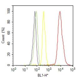
- Flow Cytometry analysis of Kindlin/FERMT1 was performed on A-431 cells labeled with ABfinity™ Anti-Kindlin/FERMT1 Recombinant Rabbit Monoclonal Antibody (Product# 701830, 2-4 ug/ 1M cells) or with Rabbit isotype control and detected with Goat anti-Rabbit IgG (H+L) Superclonal™ Secondary Antibody, (Alexa Fluor® 488 conjugate, Product # A27034, 0.4 ug/ml, 1:2500) as represented by the red and yellow histograms respectively. The purple histogram represents unstained control cells and the green histogram represents no-primary-Antibody control. A representative of 10,000 cells were acquired and analyzed for each sample using an Attune® Acoustic Focusing Cytometer (4468770).
