 Explore
 Validate
 Learn
Learn Western blot
Antibody data
- Antibody Data
- Antigen structure
- References [0]
- Comments [0]
- Validations
- Western blot [3]
- Immunocytochemistry [1]
- Immunohistochemistry [1]
Submit
Validation data
Reference
Comment
Report error
- Product number
- PA5-30002 - Provider product page

- Provider
- Invitrogen Antibodies
- Product name
- Cathepsin S Polyclonal Antibody
- Antibody type
- Polyclonal
- Antigen
- Recombinant protein fragment
- Description
- Recommended positive controls: HeLa.
- Concentration
- 1 mg/mL
No comments: Submit comment
Supportive validation
- Submitted by
- Invitrogen Antibodies (provider)
- Main image

- Experimental details
- Western Blot using Cathepsin S Polyclonal Antibody (Product # PA5-30002). Sample (30 µg of whole cell lysate). Lane A: Hela. 10% SDS PAGE. Cathepsin S Polyclonal Antibody . Cathepsin S Polyclonal Antibody (Product # PA5-30002) diluted at 1:1,000.

- Submitted by
- Invitrogen Antibodies (provider)
- Main image
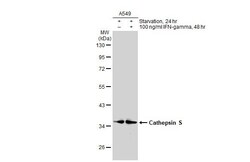
- Experimental details
- Western Blot using Cathepsin S Polyclonal Antibody (Product # PA5-30002). Untreated (–) and treated (+) A549 whole cell extracts (30 µg) were separated by 10% SDS-PAGE, and the membrane was blotted with Cathepsin S Polyclonal Antibody (Product # PA5-30002) diluted at 1:1,000. The HRP-conjugated anti-rabbit IgG antibody was used to detect the primary antibody.

- Submitted by
- Invitrogen Antibodies (provider)
- Main image

- Experimental details
- Knockdown of CTSS was achieved by transfecting A549 with CTSS specific siRNAs (Silencer® select Product # s3762, s3763). Western blot analysis (Fig. a) was performed using whole cell extracts from the CTSS knockdown cells (Lane 3), non-specific scrambled siRNA transfected cells (Lane 2) and untransfected cells (Lane 1), along with an uncharacterized band (*) at ~80 kDa. The blot was probed with Cathepsin S Polyclonal Antibody (Product # PA5-30002, 1:1000 dilution) and Rabbit anti-Goat IgG (H+L) Superclonal™ Recombinant Secondary Antibody, HRP (Product # A27014, 1:4000 dilution). Densitometric analysis of this western blot is shown in histogram (Fig. b). Decrease in signal upon siRNA mediated knock down confirms that antibody is specific to CTSS.

Supportive validation
- Submitted by
- Invitrogen Antibodies (provider)
- Main image

- Experimental details
- Immunofluorescent analysis of Cathepsin S in methanol-fixed HeLa cells using a Cathepsin S polyclonal antibody (Product # PA5-30002) (Green) at a 1:500 dilution. Alpha-tubulin filaments were labeled with Product # PA5-29281 (Red) at a 1:2000.

Supportive validation
- Submitted by
- Invitrogen Antibodies (provider)
- Main image

- Experimental details
- Cathepsin S Polyclonal Antibody detects Cathepsin S protein at lysosome on Cal27 xenograft by immunohistochemical analysis. Sample: Paraffin-embedded Cal27 xenograft. Cathepsin S Polyclonal Antibody (Product # PA5-30002) dilution: 1:500. Antigen Retrieval: EDTA based buffer, pH 8.0, 15 min.
