Explore
 Validate
 Learn
Learn Western blot
 Immunohistochemistry
Antibody data
- Antibody Data
- Antigen structure
- References [1]
- Comments [0]
- Validations
- Immunohistochemistry [1]
Submit
Validation data
Reference
Comment
Report error
- Product number
- BAF1183 - Provider product page

- Provider
- R&D Systems
- Product name
- Human Cathepsin S Biotinylated Antibody
- Antibody type
- Polyclonal
- Description
- Antigen Affinity-purified. Detects human Cathepsin S in ELISAs and Western blots . In sandwich immunoassays, less than 0.05% cross-reactivity with recombinant human Cathepsin A, -B, -C, -D, E, L, V, and -X/Z/P is observed.
- Reactivity
- Human
- Host
- Goat
- Conjugate
- Biotin
- Antigen sequence
P25774- Isotype
- IgG
- Vial size
- 50 ug
- Concentration
- LYOPH
- Storage
- Use a manual defrost freezer and avoid repeated freeze-thaw cycles. 12 months from date of receipt, -20 to -70 °C as supplied. 1 month, 2 to 8 °C under sterile conditions after reconstitution. 6 months, -20 to -70 °C under sterile conditions after reconstitution.
Submitted references A multiplex immunoassay for human adipokine profiling.
Schipper HS, de Jager W, van Dijk ME, Meerding J, Zelissen PM, Adan RA, Prakken BJ, Kalkhoven E
Clinical chemistry 2010 Aug;56(8):1320-8
Clinical chemistry 2010 Aug;56(8):1320-8
No comments: Submit comment
Supportive validation
- Submitted by
- R&D Systems (provider)
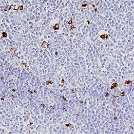
- Main image

- Experimental details
- Cathepsin S in Human Lymph Node. Cathepsin S was detected in immersion fixed paraffin-embedded sections of human lymph node using Goat Anti-Human Cathepsin S Biotinylated Antigen Affinity-purified Polyclonal Antibody (Catalog # BAF1183) at 15 µg/mL overnight at 4 °C. Tissue was stained using the Anti-Goat HRP-DAB Cell & Tissue Staining Kit (brown; Catalog # CTS008) and counterstained with hematoxylin (blue). Specific staining was localized to cytoplasm in lymphocytes. View our protocol for Chromogenic IHC Staining of Paraffin-embedded Tissue Sections.