Explore
 Validate
 Learn
Learn Western blot
Antibody data
- Antibody Data
- Antigen structure
- References [0]
- Comments [0]
- Validations
- Western blot [3]
- Immunocytochemistry [1]
- Immunohistochemistry [1]
- Flow cytometry [1]
Submit
Validation data
Reference
Comment
Report error
- Product number
- PA5-23615 - Provider product page

- Provider
- Invitrogen Antibodies
- Product name
- Fetuin A Polyclonal Antibody
- Antibody type
- Polyclonal
- Antigen
- Synthetic peptide
- Reactivity
- Human
- Host
- Rabbit
- Isotype
- IgG
- Vial size
- 400 µL
- Storage
- Store at 4°C short term. For long term storage, store at -20°C, avoiding freeze/thaw cycles.
No comments: Submit comment
Supportive validation
- Submitted by
- Invitrogen Antibodies (provider)
- Main image

- Experimental details
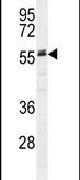
- Western blot analysis using an AHSG polyclonal antibody (Product # PA5-23615) in HepG2 cell lysates (35 µg per lane).
- Submitted by
- Invitrogen Antibodies (provider)
- Main image

- Experimental details
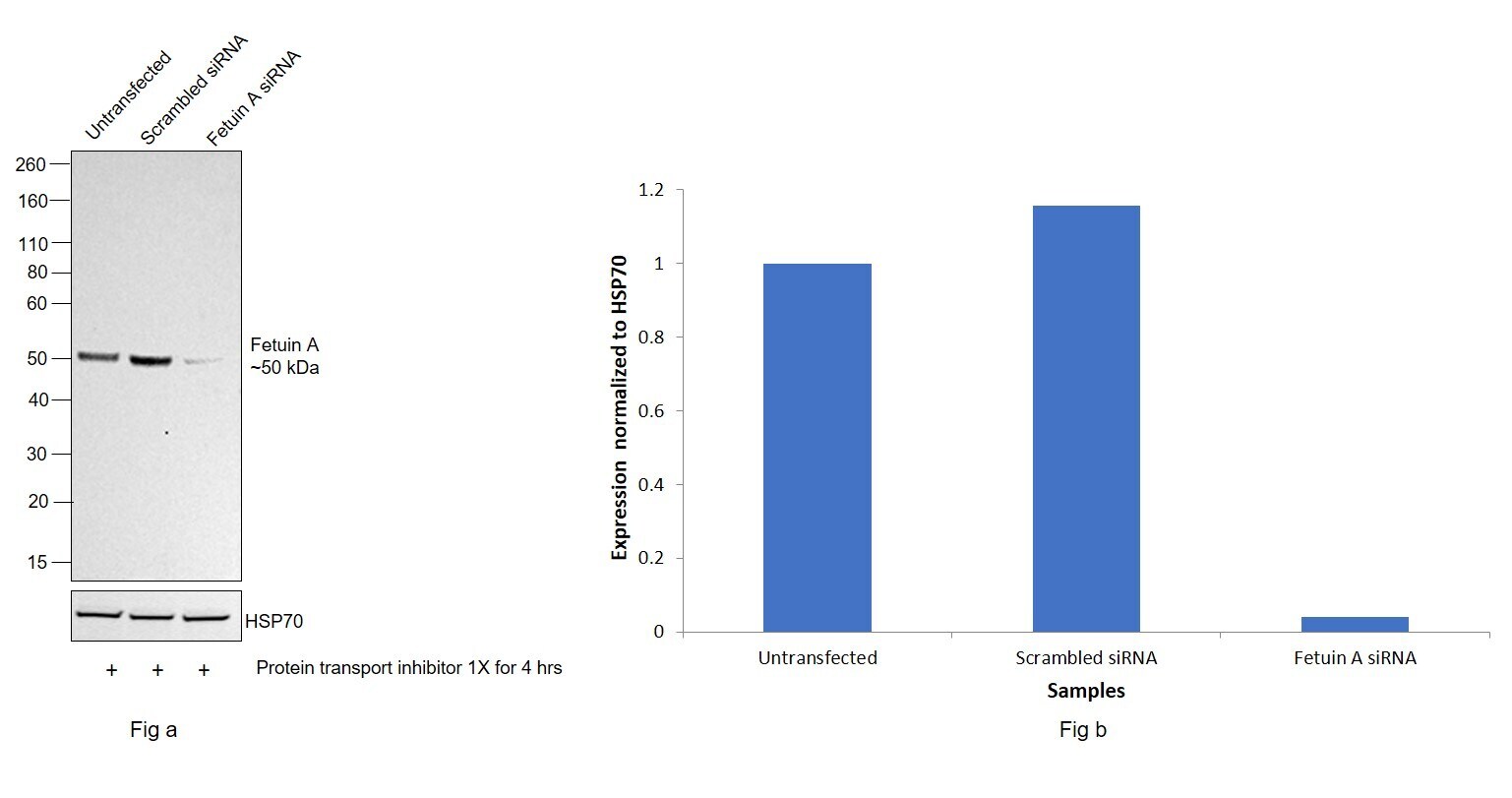
- Knockdown of Fetuin A was achieved by transfecting Hep G2 cells treated with Protein Transport Inhibitor with Fetuin A specific siRNAs (Silencer® select Product # S1201, S1203). Western blot analysis (Fig. a) was performed using whole cell extracts from the Fetuin A knockdown cells (lane 3), non-targeting scrambled siRNA transfected cells (lane 2), and untransfected cells (lane 1). The blot was probed with Fetuin A Polyclonal Antibody (Product # PA5-23615, 1:1000) and Goat anti-Rabbit IgG (H+L) Superclonal™ Recombinant Secondary Antibody, HRP (Product # A27036, 1:4000 dilution). Densitometric analysis of this western blot is shown in the histogram (Fig. b). A decrease in signal upon siRNA mediated knock down confirms that antibody is specific to Fetuin A.
- Submitted by
- Invitrogen Antibodies (provider)
- Main image
- Experimental details
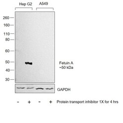
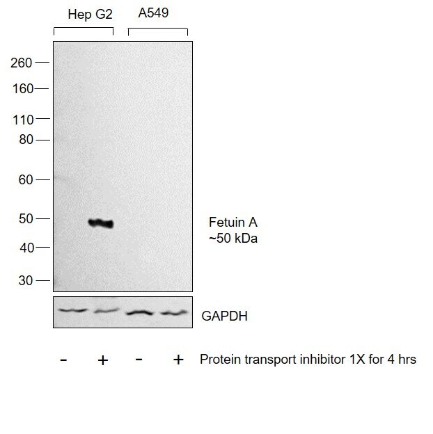
- Western blot was performed using Anti-Fetuin A Polyclonal Antibody (Product # PA5-23615) and 50 kDa band corresponding to Fetuin A (AHSG) was observed in Hep G2 treated cells but not in A549 cells as reported in literature. whole cell extracts (30 µg lysate) of Hep G2 (Lane 1), Hep G2 treated with Protein Transport Inhibitor (1X) for 4hrs (Lane 2), A549 (Lane 3), and A549 treated with Protein Transport Inhibitor (1X) for 4hrs (Lane 4) were electrophoresed using NuPAGE™ 4-12% Bis-Tris Protein Gel (Product # NP0322BOX). Resolved proteins were then transferred onto a nitrocellulose membrane (Product # IB23001) by iBlot® 2 Dry Blotting System (Product # IB21001). The blot was probed with the primary antibody (1:1000 dilution) and detected by chemiluminescence with Goat anti-Rabbit IgG (H+L) Superclonal™ Recombinant Secondary Antibody, HRP (Product # A27036, 1:4000 dilution) using the iBright FL 1000 (Product # A32752). Chemiluminescent detection was performed using Novex® ECL Chemiluminescent Substrate Reagent Kit (Product # WP20005).
Supportive validation
- Submitted by
- Invitrogen Antibodies (provider)
- Main image

- Experimental details
- Immunofluorescent analysis of HepG2 cells using an AHSG polyclonal antibody (Product # PA5-23615) at a dilution of 1:10-50, followed by a fluor-conjugated goat anti-rabbit secondary antibody (green). Nuclei were stained with DAPI (blue).

Supportive validation
- Submitted by
- Invitrogen Antibodies (provider)
- Main image

- Experimental details
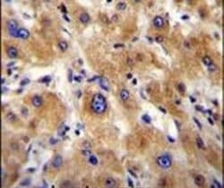
- Immunohistochemistry analysis in formalin-fixed, paraffin-embedded human hepatocarcinoma using an AHSG polyclonal antibody (Product # PA5-23615), followed by HRP-conjugated secondary antibody and DAB staining.
Supportive validation
- Submitted by
- Invitrogen Antibodies (provider)
- Main image

- Experimental details
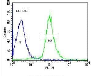
- Flow cytometry analysis of HepG2 cells using an AHSG polyclonal antibody (Product # PA5-23615) (right) compared to a negative control cell (left) at a dilution of 1:10-50, followed by a FITC-conjugated goat anti-rabbit antibody