Explore
 Validate
 Learn
Learn Western blot
Antibody data
- Antibody Data
- Antigen structure
- References [1]
- Comments [0]
- Validations
- Western blot [1]
- Other assay [1]
Submit
Validation data
Reference
Comment
Report error
- Product number
- MA1-43027 - Provider product page

- Provider
- Invitrogen Antibodies
- Product name
- SPARC Monoclonal Antibody (5031)
- Antibody type
- Monoclonal
- Antigen
- Other
- Description
- MA1-43027 detects SPARC in human, rat and bovine samples.
- Antibody clone number
- 5031
- Concentration
- 9.7 mg/mL
Submitted references Generation of Osteosarcomas from a Combination of Rb Silencing and c-Myc Overexpression in Human Mesenchymal Stem Cells.
Wang JY, Wu PK, Chen PC, Lee CW, Chen WM, Hung SC
Stem cells translational medicine 2017 Feb;6(2):512-526
Stem cells translational medicine 2017 Feb;6(2):512-526
No comments: Submit comment
Supportive validation
- Submitted by
- Invitrogen Antibodies (provider)
- Main image
- Experimental details
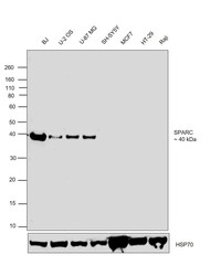
- Western blot was performed using Anti-SPARC Monoclonal Antibody (5031) (Product # MA1-43027) and a 40 kDa band corresponding to SPARC was observed in BJ, U-2 OS, U-87 MG and SH-SY5Y which are reported to be positive and not in other cell lines like MCF7, HT-29 and Raji. Whole cell extracts (30 µg lysate) of BJ (Lane 1), U-2 OS (Lane 2), U-87 MG (Lane 3), SH-SY5Y (Lane 4), MCF7 (Lane 5), HT-29 (Lane 6) and Raji (Lane 7) were electrophoresed using NuPAGE™ 10% Bis-Tris Protein Gel (Product # NP0302BOX). Resolved proteins were then transferred onto a Nitrocellulose membrane (Product # IB23001) by iBlot® 2 Dry Blotting System (Product # IB21001). The blot was probed with the primary antibody (1:1000 dilution) and detected by chemiluminescence with Goat anti-Rabbit IgG (H+L) Superclonal™ Recombinant Secondary Antibody, HRP (Product # A27036,1:4000 dilution) using the iBright FL 1000 (Product # A32752). Chemiluminescent detection was performed using Novex® ECL Chemiluminescent Substrate Reagent Kit (Product # WP20005).

Supportive validation
- Submitted by
- Invitrogen Antibodies (provider)
- Main image

- Experimental details
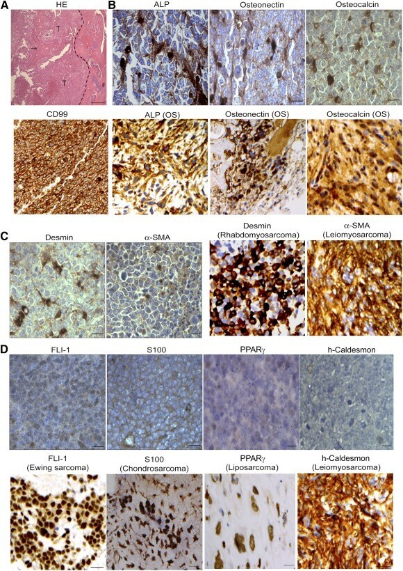
- Figure 4 Tumors formed by retinoblastoma SiRb-OeMyc express OS features. (A): HE staining of intraosseous tumor formed by SiRb-OeMyc showed new bone formation (arrow) within tumor. Dotted line indicates the tumor margin. (B-D): Immunohistochemistry shows tumors formed by SiRb-OeMyc are positive for OS markers such as CD99, ALP, osteonectin, and osteocalcin (B , upper panels; brown ) ; and slightly positive for sarcoma markers such as desmin and alpha-SMA (C , left panels; brown ) ; but negative for markers of Ewing sarcoma (FLI-1), chondrosarcoma (S100), liposarcoma (PPARgamma), and leiomyosarcoma (h-caldesmon) (D) . Positive controls of immunohistochemistry are from human pathological sections of OS (B , lower panels ) , rhabdomyosarcoma, leiomyosarcoma (C , right panels ) , Ewing sarcoma, chondrosarcoma, liposarcoma, and leiomyosarcoma (D , upper panels ) . Bars = 100 mum. Abbreviations: ALP, alkaline phosphatase; HE, hematoxylin and eosin; OS, osteosarcoma; PPARgamma, peroxisome proliferator-activated receptor gamma; alpha-SMA, alpha-smooth muscle actin; T, tumor.