Explore
 Validate
 Learn
Learn Western blot
 Flow cytometry
Antibody data
- Antibody Data
- Antigen structure
- References [8]
- Comments [0]
- Validations
- Western blot [2]
- ELISA [1]
- Immunohistochemistry [1]
Submit
Validation data
Reference
Comment
Report error
- Product number
- AF941 - Provider product page

- Provider
- R&D Systems
- Product name
- Human SPARC Antibody
- Antibody type
- Polyclonal
- Description
- Antigen Affinity-purified. Detects human SPARC/Osteonectin in direct ELISAs and Western blots. In direct ELISAs, approximately 25% cross-reactivity with recombinant mouse SPARC is observed, and less than 3% cross-reactivity with recombinant human SPARC L1 is observed.
- Reactivity
- Human
- Host
- Goat
- Conjugate
- Unconjugated
- Antigen sequence
P09486- Isotype
- IgG
- Vial size
- 100 ug
- Concentration
- LYOPH
- Storage
- Use a manual defrost freezer and avoid repeated freeze-thaw cycles. 12 months from date of receipt, -20 to -70 °C as supplied. 1 month, 2 to 8 °C under sterile conditions after reconstitution. 6 months, -20 to -70 °C under sterile conditions after reconstitution.
Submitted references In-depth proteomic analysis of tissue interstitial fluid for hepatocellular carcinoma serum biomarker discovery.
Stabilin-1 is expressed in human breast cancer and supports tumor growth in mammary adenocarcinoma mouse model.
Automated Analysis and Classification of Histological Tissue Features by Multi-Dimensional Microscopic Molecular Profiling.
Collagen signaling enhances tumor progression after anti-VEGF therapy in a murine model of pancreatic ductal adenocarcinoma.
SPARC is a key regulator of proliferation, apoptosis and invasion in human ovarian cancer.
Targeting SPARC by lentivirus-mediated RNA interference inhibits cervical cancer cell growth and metastasis.
Diverse functions of reactive cysteines facilitate unique biosynthetic processes of aggregate-prone interleukin-31.
SPARC is a VCAM-1 counter-ligand that mediates leukocyte transmigration.
Zhang J, Hao N, Liu W, Lu M, Sun L, Chen N, Wu M, Zhao X, Xing B, Sun W, He F
British journal of cancer 2017 Nov 21;117(11):1676-1684
British journal of cancer 2017 Nov 21;117(11):1676-1684
Stabilin-1 is expressed in human breast cancer and supports tumor growth in mammary adenocarcinoma mouse model.
Riabov V, Yin S, Song B, Avdic A, Schledzewski K, Ovsiy I, Gratchev A, Llopis Verdiell M, Sticht C, Schmuttermaier C, Schönhaber H, Weiss C, Fields AP, Simon-Keller K, Pfister F, Berlit S, Marx A, Arnold B, Goerdt S, Kzhyshkowska J
Oncotarget 2016 May 24;7(21):31097-110
Oncotarget 2016 May 24;7(21):31097-110
Automated Analysis and Classification of Histological Tissue Features by Multi-Dimensional Microscopic Molecular Profiling.
Riordan DP, Varma S, West RB, Brown PO
PloS one 2015;10(7):e0128975
PloS one 2015;10(7):e0128975
Collagen signaling enhances tumor progression after anti-VEGF therapy in a murine model of pancreatic ductal adenocarcinoma.
Aguilera KY, Rivera LB, Hur H, Carbon JG, Toombs JE, Goldstein CD, Dellinger MT, Castrillon DH, Brekken RA
Cancer research 2014 Feb 15;74(4):1032-44
Cancer research 2014 Feb 15;74(4):1032-44
SPARC is a key regulator of proliferation, apoptosis and invasion in human ovarian cancer.
Chen J, Wang M, Xi B, Xue J, He D, Zhang J, Zhao Y
PloS one 2012;7(8):e42413
PloS one 2012;7(8):e42413
Targeting SPARC by lentivirus-mediated RNA interference inhibits cervical cancer cell growth and metastasis.
Chen J, Shi D, Liu X, Fang S, Zhang J, Zhao Y
BMC cancer 2012 Oct 10;12:464
BMC cancer 2012 Oct 10;12:464
Diverse functions of reactive cysteines facilitate unique biosynthetic processes of aggregate-prone interleukin-31.
Shen M, Siu S, Byrd S, Edelmann KH, Patel N, Ketchem RR, Mehlin C, Arnett HA, Hasegawa H
Experimental cell research 2011 Apr 15;317(7):976-93
Experimental cell research 2011 Apr 15;317(7):976-93
SPARC is a VCAM-1 counter-ligand that mediates leukocyte transmigration.
Kelly KA, Allport JR, Yu AM, Sinh S, Sage EH, Gerszten RE, Weissleder R
Journal of leukocyte biology 2007 Mar;81(3):748-56
Journal of leukocyte biology 2007 Mar;81(3):748-56
No comments: Submit comment
Supportive validation
- Submitted by
- R&D Systems (provider)
- Main image

- Experimental details
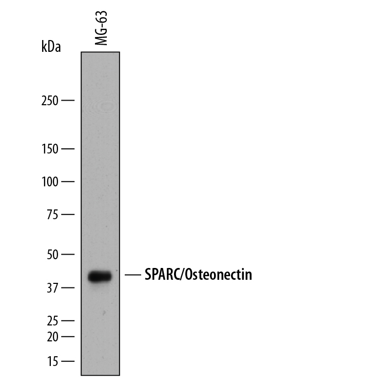
- Detection of Human SPARC by Western Blot. Western blot shows lysates of MG-63 human osteosarcoma cell line. PVDF membrane was probed with 2 µg/mL of Goat Anti-Human SPARC Antigen Affinity-purified Polyclonal Antibody (Catalog # AF941) followed by HRP-conjugated Anti-Goat IgG Secondary Antibody (Catalog # HAF019). A specific band was detected for SPARC at approximately 43 kDa (as indicated). This experiment was conducted under reducing conditions and using Immunoblot Buffer Group 1.
- Submitted by
- R&D Systems (provider)
- Main image

- Experimental details
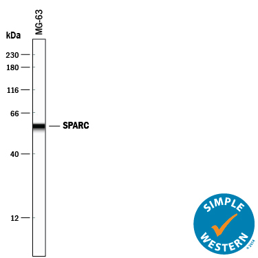
- Detection of Human SPARC by Simple WesternTM. Simple Western lane view shows lysates of MG-63 human osteosarcoma cell line, loaded at 0.2 mg/mL. A specific band was detected for SPARC at approximately 57 kDa (as indicated) using 20 µg/mL of Goat Anti-Human SPARC Antigen Affinity-purified Polyclonal Antibody (Catalog # AF941) followed by 1:50 dilution of HRP-conjugated Anti-Goat IgG Secondary Antibody (Catalog # HAF109). This experiment was conducted under reducing conditions and using the 12-230 kDa separation system.
Supportive validation
- Submitted by
- R&D Systems (provider)
- Main image

- Experimental details
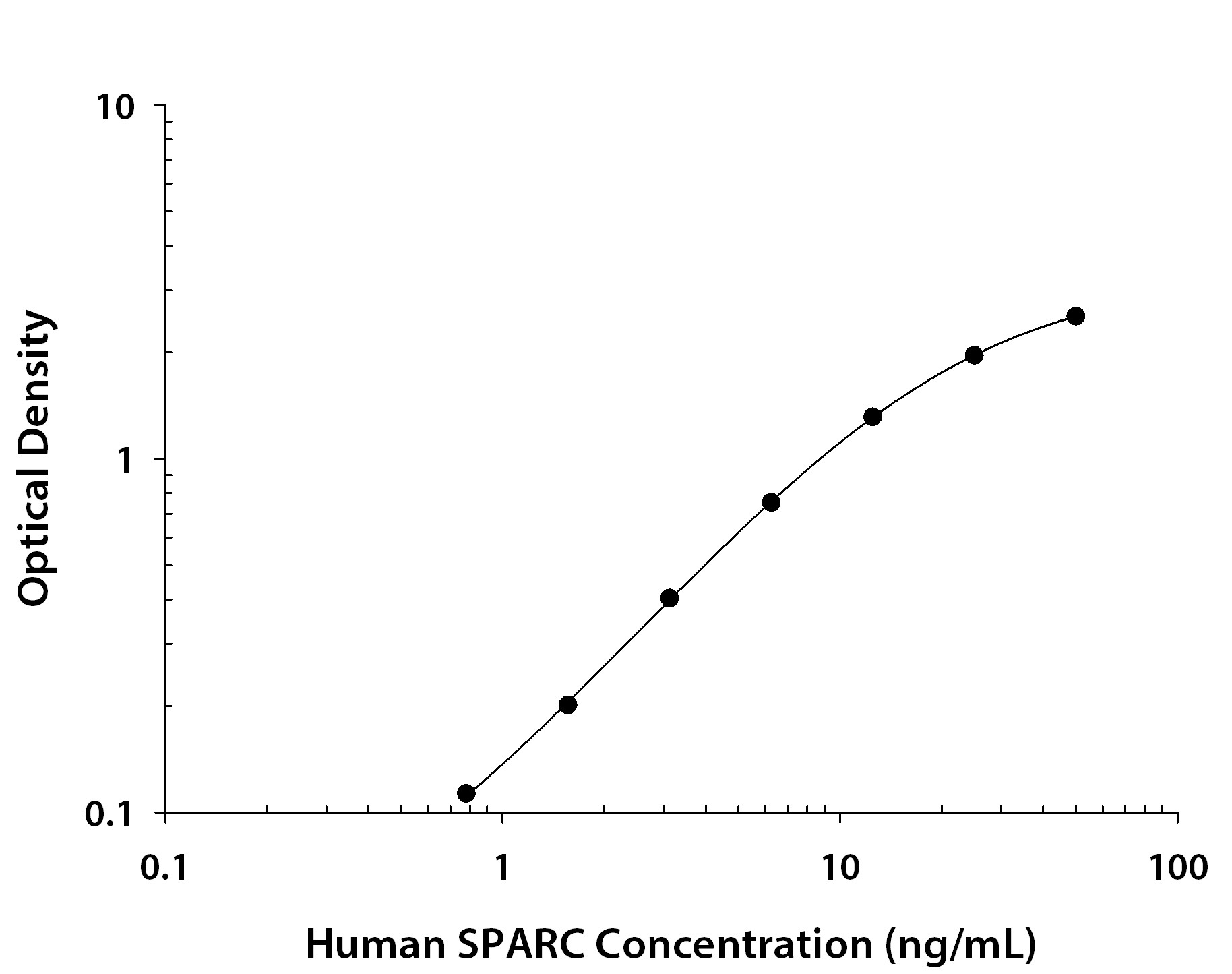
- Human SPARC ELISA Standard Curve. Recombinant Human SPARC protein was serially diluted 2-fold and captured by Mouse Anti-Human SPARC Monoclonal Antibody (Catalog # MAB941) coated on a Clear Polystyrene Microplate (Catalog # DY990). Goat Anti-Human SPARC Antigen Affinity-purified Polyclonal Antibody (Catalog # AF941) was biotinylated and incubated with the protein captured on the plate. Detection of the standard curve was achieved by incubating Streptavidin-HRP (Catalog # DY998) followed by Substrate Solution (Catalog # DY999) and stopping the enzymatic reaction with Stop Solution (Catalog # DY994).
Supportive validation
- Submitted by
- R&D Systems (provider)
- Main image

- Experimental details
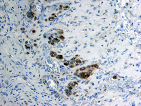
- SPARC/Osteonectin in Human Ovary. SPARC/Osteonectin was detected in immersion fixed paraffin-embedded sections of human ovary using Goat Anti-Human SPARC/Osteonectin Antigen Affinity-purified Polyclonal Antibody (Catalog # AF941) at 3 µg/mL overnight at 4 °C. Tissue was stained using the Anti-Goat HRP-DAB Cell & Tissue Staining Kit (brown; Catalog # CTS008) and counterstained with hematoxylin (blue). View our protocol for Chromogenic IHC Staining of Paraffin-embedded Tissue Sections.