Antibody data
- Antibody Data
- Antigen structure
- References [6]
- Comments [0]
- Validations
- Western blot [3]
- Immunohistochemistry [1]
Submit
Validation data
Reference
Comment
Report error
- Product number
- AF1014 - Provider product page

- Provider
- R&D Systems
- Product name
- Human Cathepsin D Antibody
- Antibody type
- Polyclonal
- Description
- Antigen Affinity-purified. Detects human Cathepsin D in direct ELISAs and Western blots. In direct ELISAs, approximately 20% cross-reactivity with recombinant mouse (rm) Cathepsin D is observed.
- Reactivity
- Human
- Host
- Goat
- Conjugate
- Unconjugated
- Antigen sequence
P07339- Isotype
- IgG
- Vial size
- 100 ug
- Concentration
- LYOPH
- Storage
- Use a manual defrost freezer and avoid repeated freeze-thaw cycles. 12 months from date of receipt, -20 to -70 °C as supplied. 1 month, 2 to 8 °C under sterile conditions after reconstitution. 6 months, -20 to -70 °C under sterile conditions after reconstitution.
Submitted references Visualizing the cellular route of entry of a cystine-knot peptide with Xfect transfection reagent by electron microscopy.
HSP90 inhibition targets autophagy and induces a CASP9-dependent resistance mechanism in NSCLC.
Amphiphilic star PEG-Camptothecin conjugates for intracellular targeting.
The Apaf-1-binding protein Aven is cleaved by Cathepsin D to unleash its anti-apoptotic potential.
IFN-gamma regulation of vacuolar pH, cathepsin D processing and autophagy in mammary epithelial cells.
Clathrin is a key regulator of basolateral polarity.
Gao X, De Mazière A, Iaea DB, Arthur CP, Klumperman J, Ciferri C, Hannoush RN
Scientific reports 2019 May 6;9(1):6907
Scientific reports 2019 May 6;9(1):6907
HSP90 inhibition targets autophagy and induces a CASP9-dependent resistance mechanism in NSCLC.
Han J, Goldstein LA, Hou W, Chatterjee S, Burns TF, Rabinowich H
Autophagy 2018;14(6):958-971
Autophagy 2018;14(6):958-971
Amphiphilic star PEG-Camptothecin conjugates for intracellular targeting.
Omar R, Bardoogo YL, Corem-Salkmon E, Mizrahi B
Journal of controlled release : official journal of the Controlled Release Society 2017 Jul 10;257:76-83
Journal of controlled release : official journal of the Controlled Release Society 2017 Jul 10;257:76-83
The Apaf-1-binding protein Aven is cleaved by Cathepsin D to unleash its anti-apoptotic potential.
Melzer IM, Fernández SB, Bösser S, Lohrig K, Lewandrowski U, Wolters D, Kehrloesser S, Brezniceanu ML, Theos AC, Irusta PM, Impens F, Gevaert K, Zörnig M
Cell death and differentiation 2012 Sep;19(9):1435-45
Cell death and differentiation 2012 Sep;19(9):1435-45
IFN-gamma regulation of vacuolar pH, cathepsin D processing and autophagy in mammary epithelial cells.
Khalkhali-Ellis Z, Abbott DE, Bailey CM, Goossens W, Margaryan NV, Gluck SL, Reuveni M, Hendrix MJ
Journal of cellular biochemistry 2008 Sep 1;105(1):208-18
Journal of cellular biochemistry 2008 Sep 1;105(1):208-18
Clathrin is a key regulator of basolateral polarity.
Deborde S, Perret E, Gravotta D, Deora A, Salvarezza S, Schreiner R, Rodriguez-Boulan E
Nature 2008 Apr 10;452(7188):719-23
Nature 2008 Apr 10;452(7188):719-23
No comments: Submit comment
Supportive validation
- Submitted by
- R&D Systems (provider)
- Main image

- Experimental details
- Detection of Human Cathepsin D by Simple Western<sup abp="263">TM. Simple Western lane view shows lysates of K562 human chronic myelogenous leukemia cell line and MCF-7 human breast cancer cell line, loaded at 0.2 mg/mL. Specific bands were detected for Procathepsin D at approximately 55 kDa and Cathepsin D heavy chain at approximately 37 kDa (as indicated) using 50 µg/mL of Goat Anti-Human Cathepsin D Antigen Affinity-purified Polyclonal Antibody (Catalog # AF1014) followed by 1:50 dilution of HRP-conjugated Anti-Goat IgG Secondary Antibody (Catalog # HAF109). This experiment was conducted under reducing conditions and using the 12-230 kDa separation system. Non-specific interaction with the 230 kDa Simple Western standard may be seen with this antibody.
- Submitted by
- R&D Systems (provider)
- Main image

- Experimental details
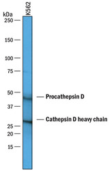
- Detection of Human Cathepsin D by Western Blot. Western blot shows lysates of K562 human chronic myelogenous leukemia cell line. PVDF membrane was probed with 1 µg/mL of Goat Anti-Human Cathepsin D Antigen Affinity-purified Polyclonal Antibody (Catalog # AF1014) followed by HRP-conjugated Anti-Goat IgG Secondary Antibody (Catalog # HAF019). Specific bands were detected for Procathepsin D at approximately 45 kDa and Cathepsin D heavy chain 28 kDa (as indicated). This experiment was conducted under reducing conditions and using Immunoblot Buffer Group 1.
- Submitted by
- R&D Systems (provider)
- Main image

- Experimental details
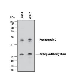
- Detection of Human Cathepsin D by Western Blot. Western blot shows lysates of PANC-1 human pancreatic carcinoma cell line and MCF-7 human breast cancer cell line. PVDF membrane was probed with 1 µg/mL of Goat Anti-Human Cathepsin D Antigen Affinity-purified Polyclonal Antibody (Catalog # AF1014) followed by HRP-conjugated Anti-Goat IgG Secondary Antibody (Catalog # HAF017). Specific bands were detected for Procathepsin D at approximately 45 kDa and Cathepsin D heavy chain 28 kDa (as indicated). This experiment was conducted under reducing conditions and using Immunoblot Buffer Group 1.
Supportive validation
- Submitted by
- R&D Systems (provider)
- Main image

- Experimental details
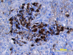
- Cathepsin D in Human Lung Cancer Tissue. Cathepsin D was detected in immersion fixed paraffin-embedded sections of human lung cancer tissue using Goat Anti-Human Cathepsin D Antigen Affinity-purified Polyclonal Antibody (Catalog # AF1014) at 15 µg/mL overnight at 4 °C. Tissue was stained using the Anti-Goat HRP-DAB Cell & Tissue Staining Kit (brown; Catalog # CTS008) and counterstained with hematoxylin (blue). View our protocol for Chromogenic IHC Staining of Paraffin-embedded Tissue Sections.
 Explore
 Validate
 Learn
Learn Western blot
 Immunoprecipitation