 Explore
 Validate
 Learn
Learn Western blot
 Immunohistochemistry
Antibody data
- Antibody Data
- Antigen structure
- References [0]
- Comments [0]
- Validations
- Western blot [3]
- Immunohistochemistry [3]
Submit
Validation data
Reference
Comment
Report error
- Product number
- HPA002131 - Provider product page

- Provider
- Atlas Antibodies
- Proper citation
- Atlas Antibodies Cat#HPA002131, RRID:AB_1080570
- Product name
- Anti-VCL
- Antibody type
- Polyclonal
- Reactivity
- Human, Mouse, Rat
- Host
- Rabbit
- Conjugate
- Unconjugated
- Antigen sequence
KMIDERQQELTHQEHRVMLVNSMNTVKELLPVLIS
AMKIFVTTKNSKNQGIEEALKNRNFTVEKMSAEIN
EIIRVLQLTSWDEDAWASKDTEAMKRALASIDSKL
NQAKGWLRDPSASPGDAGEQAIRQILDEAGKVGEL
CAGKERREI- Isotype
- IgG
- Vial size
- 100 µl
- Storage
- Store at +4°C for short term storage. Long time storage is recommended at -20°C.
No comments: Submit comment
Supportive validation
- Submitted by
- Atlas Antibodies (provider)
- Enhanced method
- Genetic validation
- Main image

- Experimental details
- Western blot analysis in U-87MG ATCC cells transfected with control siRNA, target specific siRNA probe #1 and #2, using Anti-VCL antibody. Remaining relative intensity is presented. Loading control: Anti-GAPDH.

- Submitted by
- Atlas Antibodies (provider)
- Main image

- Experimental details
- Western blot analysis in human heart tissue.

- Submitted by
- Atlas Antibodies (provider)
- Main image

- Experimental details
- Western blot analysis in mouse cell line NIH-3T3 and rat cell line NBT-II.

Enhanced validation
Supportive validation
- Submitted by
- Atlas Antibodies (provider)
- Enhanced method
- Orthogonal validation
- Main image
- Experimental details
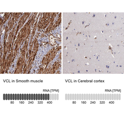
- Immunohistochemistry analysis in human smooth muscle and cerebral cortex tissues using Anti-VCL antibody. Corresponding VCL RNA-seq data are presented for the same tissues.
- Sample type
- HUMAN

Supportive validation
- Submitted by
- Atlas Antibodies (provider)
- Main image

- Experimental details
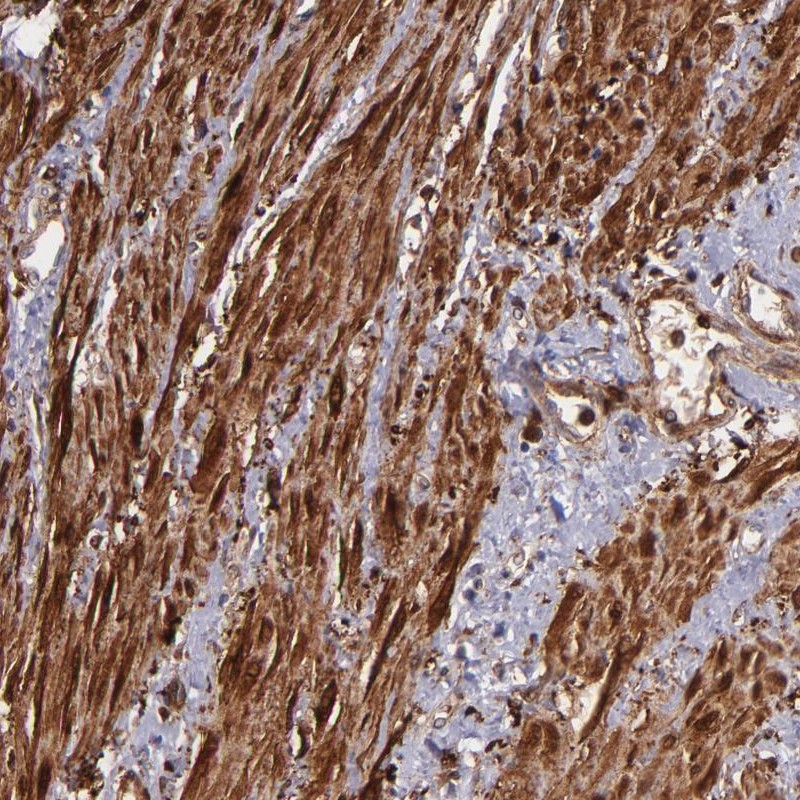
- Immunohistochemical staining of human smooth muscle shows high expression.
- Sample type
- HUMAN
- Submitted by
- Atlas Antibodies (provider)
- Main image

- Experimental details
- Immunohistochemical staining of human cerebral cortex shows low expression as expected.
- Sample type
- HUMAN
