 Explore
 Validate
 Learn
Learn Western blot
Antibody data
- Antibody Data
- Antigen structure
- References [8]
- Comments [0]
- Validations
- Western blot [7]
- Immunocytochemistry [5]
- Immunohistochemistry [5]
- Chromatin Immunoprecipitation [2]
- Other assay [8]
Submit
Validation data
Reference
Comment
Report error
- Product number
- PA5-29165 - Provider product page

- Provider
- Invitrogen Antibodies
- Product name
- SP1 Polyclonal Antibody
- Antibody type
- Polyclonal
- Antigen
- Recombinant protein fragment
- Description
- Recommended positive controls: 293T, K562, THP-1, HL-60, Raji.
- Concentration
- 0.22 mg/mL
Submitted references Treponema denticola dentilisin triggered TLR2/MyD88 activation upregulates a tissue destructive program involving MMPs via Sp1 in human oral cells.
Identification of PIM1 substrates reveals a role for NDRG1 phosphorylation in prostate cancer cellular migration and invasion.
Multi-layered epigenetic regulation of IRS2 expression in the liver of obese individuals with type 2 diabetes.
Analysis of proximal ALOX5 promoter binding proteins by quantitative proteomics.
Ischemia-induced ACSL4 activation contributes to ferroptosis-mediated tissue injury in intestinal ischemia/reperfusion.
Specific protein 1, c-Abl and ERK1/2 form a regulatory loop.
Decitabine Inhibits Gamma Delta T Cell Cytotoxicity by Promoting KIR2DL2/3 Expression.
Long interspersed nuclear element-1 expression and retrotransposition in prostate cancer cells.
Ganther S, Radaic A, Malone E, Kamarajan P, Chang NN, Tafolla C, Zhan L, Fenno JC, Kapila YL
PLoS pathogens 2021 Jul;17(7):e1009311
PLoS pathogens 2021 Jul;17(7):e1009311
Identification of PIM1 substrates reveals a role for NDRG1 phosphorylation in prostate cancer cellular migration and invasion.
Ledet RJ, Ruff SE, Wang Y, Nayak S, Schneider JA, Ueberheide B, Logan SK, Garabedian MJ
Communications biology 2021 Jan 4;4(1):36
Communications biology 2021 Jan 4;4(1):36
Multi-layered epigenetic regulation of IRS2 expression in the liver of obese individuals with type 2 diabetes.
Krause C, Geißler C, Tackenberg H, El Gammal AT, Wolter S, Spranger J, Mann O, Lehnert H, Kirchner H
Diabetologia 2020 Oct;63(10):2182-2193
Diabetologia 2020 Oct;63(10):2182-2193
Analysis of proximal ALOX5 promoter binding proteins by quantitative proteomics.
Schlag K, Steinhilber D, Karas M, Sorg BL
The FEBS journal 2020 Oct;287(20):4481-4499
The FEBS journal 2020 Oct;287(20):4481-4499
Ischemia-induced ACSL4 activation contributes to ferroptosis-mediated tissue injury in intestinal ischemia/reperfusion.
Li Y, Feng D, Wang Z, Zhao Y, Sun R, Tian D, Liu D, Zhang F, Ning S, Yao J, Tian X
Cell death and differentiation 2019 Nov;26(11):2284-2299
Cell death and differentiation 2019 Nov;26(11):2284-2299
Specific protein 1, c-Abl and ERK1/2 form a regulatory loop.
Long J, Liao G, Wang Y, Tang DD
Journal of cell science 2019 Jan 2;132(1)
Journal of cell science 2019 Jan 2;132(1)
Decitabine Inhibits Gamma Delta T Cell Cytotoxicity by Promoting KIR2DL2/3 Expression.
Niu C, Li M, Zhu S, Chen Y, Zhou L, Xu D, Li W, Cui J, Liu Y, Chen J
Frontiers in immunology 2018;9:617
Frontiers in immunology 2018;9:617
Long interspersed nuclear element-1 expression and retrotransposition in prostate cancer cells.
Briggs EM, Ha S, Mita P, Brittingham G, Sciamanna I, Spadafora C, Logan SK
Mobile DNA 2018;9:1
Mobile DNA 2018;9:1
No comments: Submit comment
Supportive validation
- Submitted by
- Invitrogen Antibodies (provider)
- Main image

- Experimental details
- Western blot analysis of SP1 using 30 µg of THP-1 lysate. Samples were loaded onto a 5% SDS-PAGE gel and probed with a SP1 polyclonal antibody (Product # PA5-29165) at a dilution of 1:2000.

- Submitted by
- Invitrogen Antibodies (provider)
- Main image

- Experimental details
- Western blot analysis of SP1 in whole cell extracts (30 µg). Samples was separated by 7.5% SDS-PAGE and the membrane was probed with SP1 Polyclonal antibody (Product # PA5-29165) at a dilution of 1:2000.

- Submitted by
- Invitrogen Antibodies (provider)
- Main image
- Experimental details
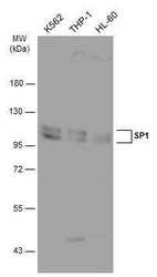
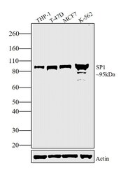
- Western Blot analysis of SP1 was performed by separating 30 µg of various whole cell extracts by 7.5% SDS-PAGE. Proteins were transferred to a membrane and probed with a SP1 Polyclonal Antibody (Product # PA5-29165) at a dilution of 1:2000 and a HRP-conjugated anti-rabbit IgG secondary antibody.
- Submitted by
- Invitrogen Antibodies (provider)
- Main image
- Experimental details
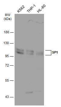
- Western Blot analysis of SP1 was performed by separating 30 µg of various whole cell extracts by 7.5% SDS-PAGE. Proteins were transferred to a membrane and probed with a SP1 Polyclonal Antibody (Product # PA5-29165) at a dilution of 1:2000 and a HRP-conjugated anti-rabbit IgG secondary antibody.

- Submitted by
- Invitrogen Antibodies (provider)
- Main image

- Experimental details
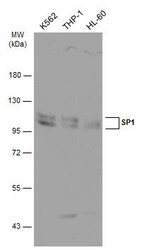
- Western Blot using SP1 Polyclonal Antibody (Product # PA5-29165). Various whole cell extracts (30 µg) were separated by 7.5% SDS-PAGE, and the membrane was blotted with SP1 Polyclonal Antibody (Product # PA5-29165) diluted at 1:2,000. The HRP-conjugated anti-rabbit IgG antibody was used to detect the primary antibody.

- Submitted by
- Invitrogen Antibodies (provider)
- Main image
- Experimental details
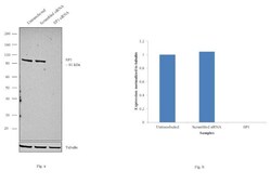
- Knockdown of SP1 was achieved by transfecting MCF7 cells with SP1 specific siRNAs (Silencer® select Product # s13319, s13320). Western blot analysis (Fig. a) was performed using whole cell extracts from the SP1 knockdown cells (lane 3), non-specific scrambled siRNA transfected cells (lane 2) and untransfected cells (lane 1). The blots were probed with SP1 Polyclonal Antibody (Product # PA5-29165, 1:1000 dilution) and Goat anti-Rabbit IgG (H+L) Superclonal™ Secondary Antibody, HRP conjugate (Product # A27036, 0.25 µg/mL, 1:4000 dilution). Densitometric analysis of this western blot is shown in histogram (Fig. b). Decrease in signal upon siRNA mediated knock down confirms that antibody is specific to SP1.

- Submitted by
- Invitrogen Antibodies (provider)
- Main image
- Experimental details
- Western blot analysis was performed on modified whole cell extracts (1% SDS) of THP-1 (Lane 1), T-47D (Lane 2), MCF7 (Lane 3) and K-562 (Lane 4). The blot was probed with SP1 Polyclonal Antibody (Product # PA5-29165, 1:1000 dilution) and detected by chemiluminescence using Goat anti-Rabbit IgG (H+L) Superclonal™ Secondary Antibody, HRP conjugate (Product # A27036, 0.25 µg/mL, 1:4000 dilution). A 95 kDa band corresponding to SP1 was observed across the panel tested.

Supportive validation
- Submitted by
- Invitrogen Antibodies (provider)
- Main image

- Experimental details
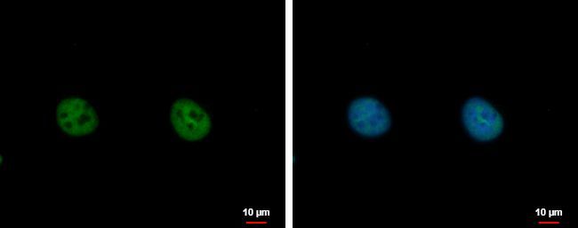
- Immunofluorescent analysis of SP1 showing staining in the nucleus of HeLa cells. HeLa cells were fixed in 4% paraformaldehyde at RT for 15 min and stained using a SP1 polyclonal antibody (Product # PA5-29165) diluted at 1:500. Blue: Hoechst 33342 staining. Scale bar = 10µm.
- Submitted by
- Invitrogen Antibodies (provider)
- Main image

- Experimental details
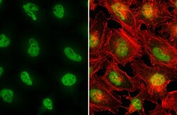
- Immunocytochemistry-Immunofluorescence analysis of SP1 was performed in HeLa cells fixed in 4% paraformaldehyde at RT for 15 min. Green: SP1 Polyclonal Antibody (Product # PA5-29165) diluted at 1:500. Red: phalloidin, a cytoskeleton marker. Scale bar = 10 µm.

- Submitted by
- Invitrogen Antibodies (provider)
- Main image
- Experimental details
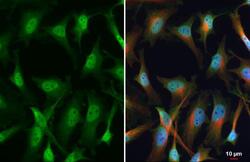
- SP1 Polyclonal Antibody detects SP1 protein at nucleus by immunofluorescent analysis. Sample: HeLa cells were fixed in 4% paraformaldehyde at RT for 15 min. Green: SP1 stained by SP1 Polyclonal Antibody (Product # PA5-29165) diluted at 1:1,000. Red: phalloidin, a cytoskeleton marker, diluted at 1:200.

- Submitted by
- Invitrogen Antibodies (provider)
- Main image
- Experimental details
- SP1 Polyclonal Antibody detects SP1 protein at nucleus by immunofluorescent analysis. Sample: HeLa cells were fixed in 4% paraformaldehyde at RT for 15 min. Green: SP1 stained by SP1 Polyclonal Antibody (Product # PA5-29165) diluted at 1:100. Red: alpha Tubulin, a cytoskeleton marker, stained by alpha Tubulin antibody [GT114] (Product # MA5-31466) diluted at 1:1,000. Blue: Fluoroshield with DAPI .

- Submitted by
- Invitrogen Antibodies (provider)
- Main image

- Experimental details
- Immunofluorescence analysis of SP1 was performed using 70% confluent log phase HeLa cells. The cells were fixed with 4% paraformaldehyde for 10 minutes, permeabilized with 0.1% Triton™ X-100 for 15 minutes, and blocked with 1% BSA for 1 hour at room temperature. The cells were labeled with SP1 Polyclonal Antibody (Product # PA5-29165) at 5 µg/mL concentration in 0.1% BSA, incubated at 4 degree Celsius overnight and then labeled with Goat anti-Rabbit IgG (H+L) Superclonal™ Secondary Antibody, Alexa Fluor® 488 conjugate (Product # A27034) for 45 minutes at room temperature (Panel a: green). Nuclei (Panel b: blue) were stained with ProLong™ Diamond Antifade Mountant with DAPI (Product # P36962). F-actin (Panel c: red) was stained with Rhodamine Phalloidin (Product # R415, 1:300). Panel d represents the merged image showing predominant Nuclear localization. Panel e represents control cells with no primary antibody to assess background. The images were captured at 60X magnification.

Supportive validation
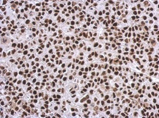
- Submitted by
- Invitrogen Antibodies (provider)
- Main image

- Experimental details
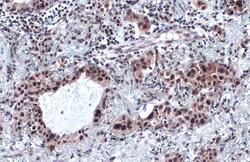
- Immunohistochemistry (Paraffin) analysis of SP1 was performed in paraffin-embedded human breast carcinoma tissue using SP1 Polyclonal Antibody (Product # PA5-29165) at a dilution of 1:500. Antigen Retrieval: Citrate buffer, pH 6.0, 15 min.

- Submitted by
- Invitrogen Antibodies (provider)
- Main image
- Experimental details
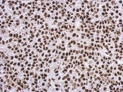
- Immunohistochemical analysis of paraffin-embedded C2C12 xenograft, using SP1 (Product # PA5-29165) antibody at 1:500 dilution. Antigen Retrieval: EDTA based buffer, pH 8.0, 15 min.
- Submitted by
- Invitrogen Antibodies (provider)
- Main image
- Experimental details
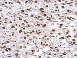
- SP1 Polyclonal Antibody detects SP1 protein at cytoplasm and nucleus by immunohistochemical analysis. Sample: Paraffin-embedded human lung cancer. SP1 stained by SP1 Polyclonal Antibody (Product # PA5-29165) diluted at 1:2,000. Antigen Retrieval: Citrate buffer, pH 6.0, 15 min.

- Submitted by
- Invitrogen Antibodies (provider)
- Main image
- Experimental details
- Immunohistochemical analysis of paraffin-embedded Hela xenograft, using SP1 (Product # PA5-29165) antibody at 1:500 dilution. Antigen Retrieval: EDTA based buffer, pH 8.0, 15 min.
- Submitted by
- Invitrogen Antibodies (provider)
- Main image

- Experimental details
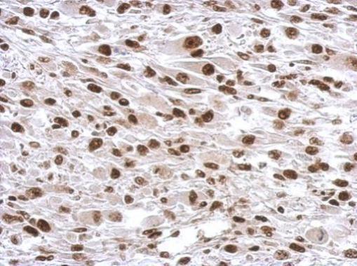
- SP1 Polyclonal Antibody detects SP1 protein at nucleus by immunohistochemical analysis. Sample: Paraffin-embedded human breast carcinoma. SP1 stained by SP1 Polyclonal Antibody (Product # PA5-29165) diluted at 1:500. Antigen Retrieval: Citrate buffer, pH 6.0, 15 min.

Supportive validation
- Submitted by
- Invitrogen Antibodies (provider)
- Main image

- Experimental details
- Cross-linked ChIP was performed with 293T chromatin extract and 5 µg of either control rabbit IgG or a SP1 polyclonal antibody (Product # PA5-29165). The precipitated DNA was detected by PCR with primer set targeting to MGARP promoter.

- Submitted by
- Invitrogen Antibodies (provider)
- Main image

- Experimental details
- ChIP assay analysis of SP1 was performed in 293T chromatin extracts using 5 µg of either normal rabbit IgG or SP1 Polyclonal Antibody (Product # PA5-29165). The precipitated DNA was detected by PCR with primer set targeting to MGARP promoter.

Supportive validation
- Submitted by
- Invitrogen Antibodies (provider)
- Main image

- Experimental details
- NULL

- Submitted by
- Invitrogen Antibodies (provider)
- Main image

- Experimental details
- NULL

- Submitted by
- Invitrogen Antibodies (provider)
- Main image

- Experimental details
- NULL

- Submitted by
- Invitrogen Antibodies (provider)
- Main image

- Experimental details
- Immunoprecipitation of SP1 was performed in THP-1 whole cell extracts using 5 µg of SP1 Polyclonal Antibody (Product # PA5-29165). Samples were transferred to a membrane and probed with SP1 Polyclonal Antibody as a primary antibody and an HRP-conjugated anti-Rabbit IgG was used as a secondary antibody.

- Submitted by
- Invitrogen Antibodies (provider)
- Main image

- Experimental details
- 10.1371/journal.ppat.1009311.g006 Fig 6 Healthy hPDL cells were challenged with A) Td-WT and B) isogenic Td-CF522 bacteria at an MOI of 50 as previously described. Whole cell lysates were generated and used for Western Blot analysis utilizing total anti-Sp1 antibodies. Total Sp1 protein expression was normalized against GAPDH protein expression as a loading control. Statistical significance was determined using a paired t-test. Bars represent mean +- SD (n = 3). **p < .01 versus control.

- Submitted by
- Invitrogen Antibodies (provider)
- Main image

- Experimental details
- Fig. 5. ERK1/2 regulates Sp1, c-Abl and cell proliferation. (A) HASM cells were treated with 10 ng/ml PDGF for 10 min in the absence or presence of 10 uM the MEK inhibitor U0126 or left unstimulated. The level of ERK1/2 phosphorylation (ERK1: Thr-202/Tyr 204; ERK2: Thr-185/Tyr-187, p-ERK1/2) was determined by immunoblot analysis. Data are mean+-s.d. ( n =4-7). * P

- Submitted by
- Invitrogen Antibodies (provider)
- Main image

- Experimental details
- 3 Fig. Validation of the experimental set-up. (A) 3% Agarose gel electrophoresis to verify annealing of forward and reverse single-stranded sequences used in the DNA pulldowns. Sequences for ALOX 5 promoter oligonucleotides (WT) and SCR controls are given in Table S1. (B) 5-LO expression pattern in myeloid cell lines HL-60, THP1, MM6 and B-lymphocytic cell lines Rec-1 and BL-41. Myeloid cells were treated with TGFbeta/1,25(OH) 2 D 3 to induce differentiation and 5-LO protein expression. Samples from three consecutive cell passages were analysed in adjacent lanes. (C) Immunoblot from DNA pulldown with nuclear extract from the B-cell line Rec-1 to verify specific binding of the transcription factor Sp1 to the ALOX5 promoter sequence (WT) as compared to SCR control. Four hundred microgram nuclear extract was used for each DNA fragment; the blotting membrane was probed with anti-Sp1 antibody (0.6 mug*mL -1 ). The result from one out of three independent repetitions is shown.

- Submitted by
- Invitrogen Antibodies (provider)
- Main image

- Experimental details
- Fig. 6 Sp1 regulates ACSL4 transcription and expression. a - c Ischemic human intestines, 45 min ischemic mouse intestines and 12 h hypoxic Caco-2 cells were used for the determination of ACSL4 mRNA levels by qPCR ( n = 3). ** p < 0.01 vs. the normal/sham/normoxia groups. d , e Expression of nuclear Sp1 at 12 h of hypoxia was determined by western blotting ( n = 3) and laser scanning confocal microscopy ( n = 6). ** p < 0.01 vs. the normoxia group. f - k Caco-2 cells were transfected with Sp1 plasmid or siRNA for 2 days, and then subjected to 12 h of hypoxia. The Sp1 protein levels, the protein and mRNA levels of ACSL4 were determined after hypoxia (n = 3). Results are expressed as the mean +- SD. * p < 0.05, ** p < 0.01 vs. the control group; # p < 0.05, ## p < 0.01 vs. the hypoxia group
