 Explore
 Validate
 Learn
Learn Flow cytometry
Antibody data
- Antibody Data
- Antigen structure
- References [17]
- Comments [0]
- Validations
- Flow cytometry [2]
- Other assay [6]
Submit
Validation data
Reference
Comment
Report error
- Product number
- 53-9320-80 - Provider product page

- Provider
- Invitrogen Antibodies
- Product name
- PLZF Monoclonal Antibody (Mags.21F7), Alexa Fluor™ 488, eBioscience™
- Antibody type
- Monoclonal
- Antigen
- Other
- Description
- Description: This Mags.21F7 monoclonal antibody reacts with human and mouse promyelocytic leukemia zinc finger (PLZF), a member of the BTB-POZ family of transcription factors. Expression of this transcriptional repressor in immune cells differs between mice and humans. In mice, PLZF is highly expressed in immature CD1d-resricted NKT2 and NKT1 cells, and a subset of gamma delta (Vg1.1+Vd6.3+) T cells. Studies have also demonstrated expression of PLZF in non-invariant CD1d-restricted T cells, as well as non-CD1d-restricted innate T cells. In humans, PLZF is expressed in NK cells, gamma delta T cells, as well as CD4 and CD8+ T cells. PLZF is also expressed in MR1-specific mucosal-associated invariant T cells, as well as in MHC Class II-restricted T cells that develop via a thymocyte-thymocyte interaction in humans. PLZF exists as a homodimer or in complex with PLZP, and has been shown to be involved in the development of NKT cells, NK cell function, cellular quiescence, and growth suppression. Finally, PLZF has been shown to inhibit gene expression induced by retinoic acid receptor. Applications Reported: This Mags.21F7 antibody has been reported for use in intracellular staining followed by flow cytometric analysis. Applications Tested: This Mags.21F7 antibody has been tested by intracellular staining followed by flow cytometric analysis of mouse thymocytes. This can be used at less than or equal to 1 µg per test. A test is defined as the amount (µg) of antibody that will stain a cell sample in a final volume of 100 µL. Cell number should be determined empirically but can range from 10^5 to 10^8 cells/test. It is recommended that the antibody be carefully titrated for optimal performance in the assay of interest. Excitation: 488 nm; Emission: 519 nm; Laser: Blue Laser. Filtration: 0.2 µm post-manufacturing filtered.
- Reactivity
- Human, Mouse
- Host
- Mouse
- Conjugate
- Green dye
- Isotype
- IgG
- Antibody clone number
- Mags.21F7
- Vial size
- 25 µg
- Concentration
- 0.5 mg/mL
- Storage
- 4° C, store in dark, DO NOT FREEZE!
Submitted references Loss of Ubiquitin Carboxy-Terminal Hydrolase L1 Impairs Long-Term Differentiation Competence and Metabolic Regulation in Murine Spermatogonial Stem Cells.
Diversity in medullary thymic epithelial cells controls the activity and availability of iNKT cells.
Cytotoxicity of nonylphenol on spermatogonial stem cells via phosphatidylinositol-3-kinase/protein kinase B/mammalian target of rapamycin pathway.
Thymic iNKT single cell analyses unmask the common developmental program of mouse innate T cells.
Transnuclear mice reveal Peyer's patch iNKT cells that regulate B-cell class switching to IgG1.
In Vitro Cytotoxicity of Folate-Silica-Gold Nanorods on Mouse Acute Lymphoblastic Leukemia and Spermatogonial Cells.
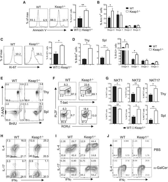
Keap1-Nrf2 System Plays an Important Role in Invariant Natural Killer T Cell Development and Homeostasis.
Stabilization of cytokine mRNAs in iNKT cells requires the serine-threonine kinase IRE1alpha.
Monoclonal Invariant NKT (iNKT) Cell Mice Reveal a Role for Both Tissue of Origin and the TCR in Development of iNKT Functional Subsets.
Distinct germline progenitor subsets defined through Tsc2-mTORC1 signaling.
Steady-state production of IL-4 modulates immunity in mouse strains and is determined by lineage diversity of iNKT cells.
Altered development of NKT cells, γδ T cells, CD8 T cells and NK cells in a PLZF deficient patient.
PLZF induces the spontaneous acquisition of memory/effector functions in T cells independently of NKT cell-related signals.
Generation of PLZF+ CD4+ T cells via MHC class II-dependent thymocyte-thymocyte interaction is a physiological process in humans.
Development of promyelocytic zinc finger and ThPOK-expressing innate gamma delta T cells is controlled by strength of TCR signaling and Id3.
The transcription factor PLZF directs the effector program of the NKT cell lineage.
PLZF is a negative regulator of retinoic acid receptor transcriptional activity.
Alpaugh WF, Voigt AL, Dardari R, Su L, Al Khatib I, Shin W, Goldsmith TM, Coyle KM, Tang LA, Shutt TE, Klein C, Biernaskie J, Dobrinski I
Cells 2021 Aug 31;10(9)
Cells 2021 Aug 31;10(9)
Diversity in medullary thymic epithelial cells controls the activity and availability of iNKT cells.
Lucas B, White AJ, Cosway EJ, Parnell SM, James KD, Jones ND, Ohigashi I, Takahama Y, Jenkinson WE, Anderson G
Nature communications 2020 May 4;11(1):2198
Nature communications 2020 May 4;11(1):2198
Cytotoxicity of nonylphenol on spermatogonial stem cells via phosphatidylinositol-3-kinase/protein kinase B/mammalian target of rapamycin pathway.
Lei JH, Yan W, Luo CH, Guo YM, Zhang YY, Wang XH, Su XJ
World journal of stem cells 2020 Jun 26;12(6):500-513
World journal of stem cells 2020 Jun 26;12(6):500-513
Thymic iNKT single cell analyses unmask the common developmental program of mouse innate T cells.
Harsha Krovi S, Zhang J, Michaels-Foster MJ, Brunetti T, Loh L, Scott-Browne J, Gapin L
Nature communications 2020 Dec 7;11(1):6238
Nature communications 2020 Dec 7;11(1):6238
Transnuclear mice reveal Peyer's patch iNKT cells that regulate B-cell class switching to IgG1.
Clancy-Thompson E, Chen GZ, LaMarche NM, Ali LR, Jeong HJ, Crowley SJ, Boelaars K, Brenner MB, Lynch L, Dougan SK
The EMBO journal 2019 Jul 15;38(14):e101260
The EMBO journal 2019 Jul 15;38(14):e101260
In Vitro Cytotoxicity of Folate-Silica-Gold Nanorods on Mouse Acute Lymphoblastic Leukemia and Spermatogonial Cells.
Eslahi N, Shakeri-Zadeh A, Ashtari K, Pirhajati-Mahabadi V, Tohidi Moghadam T, Shabani R, Kamrava K, Madjd Z, Maki C, Asgari HR, Koruji M
Cell journal 2019 Apr;21(1):14-26
Cell journal 2019 Apr;21(1):14-26
Keap1-Nrf2 System Plays an Important Role in Invariant Natural Killer T Cell Development and Homeostasis.
Pyaram K, Kumar A, Kim YH, Noel S, Reddy SP, Rabb H, Chang CH
Cell reports 2019 Apr 16;27(3):699-707.e4
Cell reports 2019 Apr 16;27(3):699-707.e4
Stabilization of cytokine mRNAs in iNKT cells requires the serine-threonine kinase IRE1alpha.
Govindarajan S, Gaublomme D, Van der Cruyssen R, Verheugen E, Van Gassen S, Saeys Y, Tavernier S, Iwawaki T, Bloch Y, Savvides SN, Lambrecht BN, Janssens S, Elewaut D, Drennan MB
Nature communications 2018 Dec 17;9(1):5340
Nature communications 2018 Dec 17;9(1):5340
Monoclonal Invariant NKT (iNKT) Cell Mice Reveal a Role for Both Tissue of Origin and the TCR in Development of iNKT Functional Subsets.
Clancy-Thompson E, Chen GZ, Tyler PM, Servos MM, Barisa M, Brennan PJ, Ploegh HL, Dougan SK
Journal of immunology (Baltimore, Md. : 1950) 2017 Jul 1;199(1):159-171
Journal of immunology (Baltimore, Md. : 1950) 2017 Jul 1;199(1):159-171
Distinct germline progenitor subsets defined through Tsc2-mTORC1 signaling.
Hobbs RM, La HM, Mäkelä JA, Kobayashi T, Noda T, Pandolfi PP
EMBO reports 2015 Apr;16(4):467-80
EMBO reports 2015 Apr;16(4):467-80
Steady-state production of IL-4 modulates immunity in mouse strains and is determined by lineage diversity of iNKT cells.
Lee YJ, Holzapfel KL, Zhu J, Jameson SC, Hogquist KA
Nature immunology 2013 Nov;14(11):1146-54
Nature immunology 2013 Nov;14(11):1146-54
Altered development of NKT cells, γδ T cells, CD8 T cells and NK cells in a PLZF deficient patient.
Eidson M, Wahlstrom J, Beaulieu AM, Zaidi B, Carsons SE, Crow PK, Yuan J, Wolchok JD, Horsthemke B, Wieczorek D, Sant'Angelo DB
PloS one 2011;6(9):e24441
PloS one 2011;6(9):e24441
PLZF induces the spontaneous acquisition of memory/effector functions in T cells independently of NKT cell-related signals.
Kovalovsky D, Alonzo ES, Uche OU, Eidson M, Nichols KE, Sant'Angelo DB
Journal of immunology (Baltimore, Md. : 1950) 2010 Jun 15;184(12):6746-55
Journal of immunology (Baltimore, Md. : 1950) 2010 Jun 15;184(12):6746-55
Generation of PLZF+ CD4+ T cells via MHC class II-dependent thymocyte-thymocyte interaction is a physiological process in humans.
Lee YJ, Jeon YK, Kang BH, Chung DH, Park CG, Shin HY, Jung KC, Park SH
The Journal of experimental medicine 2010 Jan 18;207(1):237-46
The Journal of experimental medicine 2010 Jan 18;207(1):237-46
Development of promyelocytic zinc finger and ThPOK-expressing innate gamma delta T cells is controlled by strength of TCR signaling and Id3.
Alonzo ES, Gottschalk RA, Das J, Egawa T, Hobbs RM, Pandolfi PP, Pereira P, Nichols KE, Koretzky GA, Jordan MS, Sant'Angelo DB
Journal of immunology (Baltimore, Md. : 1950) 2010 Feb 1;184(3):1268-79
Journal of immunology (Baltimore, Md. : 1950) 2010 Feb 1;184(3):1268-79
The transcription factor PLZF directs the effector program of the NKT cell lineage.
Savage AK, Constantinides MG, Han J, Picard D, Martin E, Li B, Lantz O, Bendelac A
Immunity 2008 Sep 19;29(3):391-403
Immunity 2008 Sep 19;29(3):391-403
PLZF is a negative regulator of retinoic acid receptor transcriptional activity.
Martin PJ, Delmotte MH, Formstecher P, Lefebvre P
Nuclear receptor 2003 Sep 6;1(1):6
Nuclear receptor 2003 Sep 6;1(1):6
No comments: Submit comment
Supportive validation
- Submitted by
- Invitrogen Antibodies (provider)
- Main image

- Experimental details
- C57Bl/6 thymocytes were surface-stained with Anti-Mouse TCR beta, Anti-Mouse CD1d Tetramer, and Anti-Mouse NK1-1 PerCP-Cyanine5-5 (Product # 45-5941-82), followed by intracellular staining with Anti-Human/Mouse PLZF Alexa Fluor® 488 using the Foxp3 Staining Buffer Set and protocol (Product # 00-5523-00). Anti-Human/Mouse PLZF Alexa Fluor® 488 staining (right) was compared in T cells (CD1d Tetramer-NK1-1-TCR beta+; blue histogram), NKT1 cells (CD1d Tetramer+NK1-1+TCR beta+; orange histogram), and NKT2 cells (CD1d Tetramer+NK1-1-TCR beta+; purple histogram).
- Conjugate
- Green dye

Supportive validation
- Submitted by
- Invitrogen Antibodies (provider)
- Main image

- Experimental details
- NULL
- Conjugate
- Green dye
- Submitted by
- Invitrogen Antibodies (provider)
- Main image

- Experimental details
- NULL
- Conjugate
- Green dye

- Submitted by
- Invitrogen Antibodies (provider)
- Main image

- Experimental details
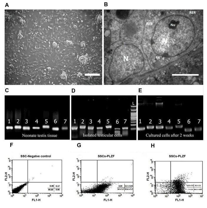
- Fig.1 Spermatogonial cells characterization. A . The morphology of a spermatogonial-derived cluster formed from the culturing of spermatogonial cells after 24 hours (scale bar: 200 um), B . Representative transmission electron micrographs from spermatogonial cells (SSCs) clusters (scale bar: 5 um). The heterochromatin nucleus (N), eccentric small compact and highly reticulated nucleoli (Nu), Rough endoplasmic reticulum (RER) and very high mitochondria (M) were observed in cells. Reverse transcription polymerase chain reaction (RT-PCR) was used to determine the expression of specific spermatogonia and germ cell markers in C . Neonate testis tissue (fresh tissue without enzymatic digestion), D . Cultured cells after the first day and E . Two weeks of culture. 1; Oct4 (129 bp), 2; Itgalpha6 (148 bp), 3; Plzf (137 bp), 4; Gfralpha1 (130 bp), 5; Mvh (Vasa, 149 bp), 6; Itgss1 (115 bp), 7; Gapdh (125 bp). Flow cytometric analysis of spermatogonial cells: F . Spermatogonial negative control, G . The PLZF positive spermatogonial cells at the end of the first week were 45.63 +- 5.71%, and H . At the end of the second week was 84.68 +- 4.02%.
- Conjugate
- Green dye
- Submitted by
- Invitrogen Antibodies (provider)
- Main image

- Experimental details
- Figure 3 Analysis of whole Uch-l1 -/- , Uch-l1 +/- and Uch-l1 +/+ testes. ( A ) Quantitative PCR results of significantly differentially expressed genes in 7, 17 and 120-day old of Uch-l1 -/- relative to Uch-l1 +/+ testes ( B , F ) Flow cytometry analysis of PLZF and c-KIT positive cells in Uch-l1 +/+ , Uch-l1 +/- and Uch-l1 -/- testes at 120 days of age. n = 6/genotype. ( B , C ) Representative dot plot of c-KIT and PLZF positive cells in Uch-l1 +/+ and Uch-l1 -/- testes, respectively. ( D ) Percentage of PLZF+ c-KIT- cells in Uch-l1 +/+ , Uch-l1 +/- and Uch-l1 -/- . ( E ) Percentage of PLZF+ c-KIT+ cells in Uch-l1 +/+ , Uch-l1 +/- and Uch-l1 -/- . ( F ) Percentage of c-KIT+ cells in Uch-l1 +/+ , Uch-l1 +/- and Uch-l1 -/- . * indicates significant differences ( p < 0.05), ** represents significant differences p < 0.01.
- Conjugate
- Green dye

- Submitted by
- Invitrogen Antibodies (provider)
- Main image

- Experimental details
- Figure 2 iNKT cells from TN and C57BL/6 mice show similar influence of tissue microenvironment on NKT1, NKT2, and NKT17 subsets A-F Lymphocytes from the indicated tissues of C57BL/6 and Valpha14 mice were stained with anti-CD3 and CD1d-(PBS57)-tetramer, before they were fixed, permeabilized, and stained with antibodies to T-bet, RORgammat, and PLZF. Results shown are gated on CD3 + CD1d-tetramer + cells. G The percentage of CD3 + CD1d-tetramer + iNKT cells in each organ that stained positively for PLZF, T-bet, and RORgammat are shown. ** P < 0.01, Mann-Whitney test. Error bars are SD. Data information: Results shown are representative of three independent experiments where n = 3 biological replicates.
- Conjugate
- Green dye

- Submitted by
- Invitrogen Antibodies (provider)
- Main image

- Experimental details
- Figure EV2 Adipose iNKT cells from Valpha14 TN mice are indistinguishable from C57 BL /6-derived adipose iNKT cells Flow cytometry analysis of iNKT cell abundance in white adipose tissue from a Valpha14 TN mouse. Spleen cells and stromal vascular fractions of white adipose tissue from Valpha14 TN or C57BL/6 mice were stained intracellularly with anti-PLZF and analyzed by flow cytometry. Histograms shown are gated on CD1d-(PBS57)-tetramer + CD3 + cells. Thymus, spleen, and adipose tissue were harvested from C57BL/6 mice and Valpha14 TN mice. Cell suspensions were stained with antibodies to CD3, Nur77, E4BP4, and CD1d-(PBS57)-tetramer and analyzed by flow cytometry. Mean fluorescence intensity of Nur77 and E4BP4 staining after gating on iNKT cells is shown. N = 3 per group. Error bars are SEM. Representative histograms of E4BP4 staining are shown. Plots are gated on total CD3 + cells.
- Conjugate
- Green dye
