Explore
 Validate
 Learn
Learn Western blot
Antibody data
- Antibody Data
- Antigen structure
- References [1]
- Comments [0]
- Validations
- Western blot [2]
- Immunohistochemistry [4]
- Other assay [3]
Submit
Validation data
Reference
Comment
Report error
- Product number
- PA5-81949 - Provider product page

- Provider
- Invitrogen Antibodies
- Product name
- PLZF Polyclonal Antibody
- Antibody type
- Polyclonal
- Antigen
- Recombinant full-length protein
- Description
- Immunogen sequence: HYTLDFLSPK TFQQILEYAY TATLQAKAED LDDLLYAAEI LEIEYLEEQC LKMLETIQAS DDNDTEATMA DGGAEEEEDR KARYLKNIFI SKHSSEESGY ASVAGQSLPG PMVDQSPSVS TSFGLSA
- Reactivity
- Human
- Host
- Rabbit
- Isotype
- IgG
- Vial size
- 100 µL
- Concentration
- 0.2 mg/mL
- Storage
- Store at 4°C short term. For long term storage, store at -20°C, avoiding freeze/thaw cycles.
Submitted references Stage-specific embryonic antigen 4 is a membrane marker for enrichment of porcine spermatogonial stem cells.
Zhang P, Li F, Zhang L, Lei P, Zheng Y, Zeng W
Andrology 2020 Nov;8(6):1923-1934
Andrology 2020 Nov;8(6):1923-1934
No comments: Submit comment
Supportive validation
- Submitted by
- Invitrogen Antibodies (provider)
- Main image
- Experimental details
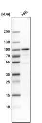
- Western blot analysis of PLZF by a PLZF polyclonal antibody (Product # PA5-81949). Analysis in human cell line HEL.

- Submitted by
- Invitrogen Antibodies (provider)
- Main image
- Experimental details
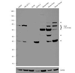
- Western blot was performed using Anti-PLZF Polyclonal Antibody (Product # PA5-81949) and a 74 kDa band corresponding to ZBTB16 was observed across positive cell lines LNCaP, HEL 92.1.7 and mouse tissue lysates but not in negative cell lines HL-60 and SK-OV-3. Tissue lysates also detect the second isoform of PLZF (~61 kDa). Nuclear enriched extracts (30 µg lysate) of LNCaP (Lane 1), HEL 92.1.7 (Lane 2), HL-60 (Lane 3), SK-O-V3 (Lane 4), Mouse Heart (Lane 5), Mouse Ovary (Lane 6) and Mouse Thymus (Lane 7) were electrophoresed using NuPAGE™ 4-12% Bis-Tris Protein Gel (Product # NP0322BOX). Resolved proteins were then transferred onto a nitrocellulose membrane (Product # IB23001) by iBlot® 2 Dry Blotting System (Product # IB21001). The blot was probed with the primary antibody (0.3 µg/mL dilution) and detected by chemiluminescence with Goat anti-Rabbit IgG (H+L) Superclonal™ Recombinant Secondary Antibody, HRP (Product # A27036,1:10000 dilution) using the iBright™ FL1500 Imaging System (Product # A44115). Chemiluminescent detection was performed using SuperSignal™ West Pico PLUS Chemiluminescent Substrate (Product # 34580). An uncharacterized band (*) was observed at ~40 kDa across cell and tissue lysates except in HL-60 and Mouse Heart. Additionally, the tissue lysates display two other uncharacterized bands (*) at ~55 kDa and ~80 kDa.

Supportive validation
- Submitted by
- Invitrogen Antibodies (provider)
- Main image
- Experimental details
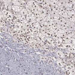
- Immunohistochemical analysis of PLZF in human adrenal gland using a PLZF polyclonal antibody (Product # PA5-81949). The analysis shows moderate to strong nuclear positivity in glandular cells.
- Submitted by
- Invitrogen Antibodies (provider)
- Main image

- Experimental details
- Immunohistochemical analysis of PLZF in human cerebral cortex using a PLZF polyclonal antibody (Product # PA5-81949). The analysis shows moderate to strong nuclear positivity in neuronal cells.

- Submitted by
- Invitrogen Antibodies (provider)
- Main image
- Experimental details
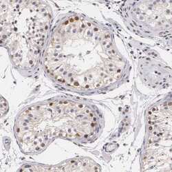
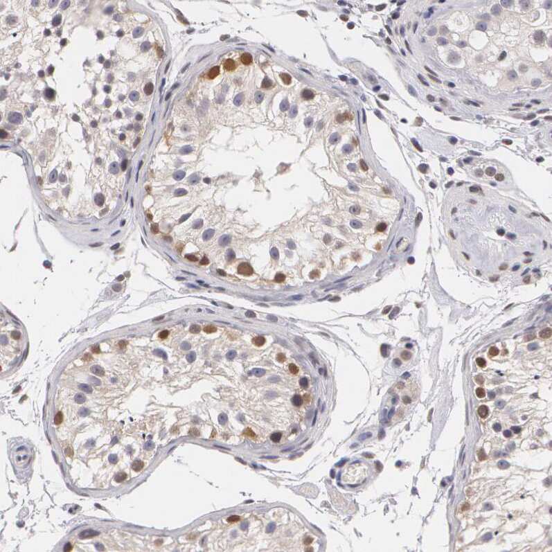
- Immunohistochemical analysis of PLZF in human testis using a PLZF polyclonal antibody (Product # PA5-81949). The analysis shows moderate to strong positivity in nuclear membrane in cells in seminiferous ducts.
- Submitted by
- Invitrogen Antibodies (provider)
- Main image

- Experimental details
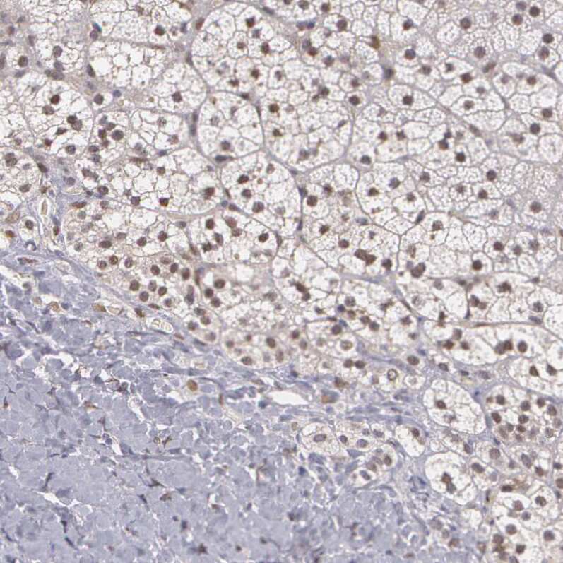
- Immunohistochemical analysis of PLZF in human kidney using a PLZF polyclonal antibody (Product # PA5-81949). The analysis shows no positivity in cells in tubules as expected.

Supportive validation
- Submitted by
- Invitrogen Antibodies (provider)
- Main image

- Experimental details
- 2 FIGURE Co-immunostaining of the SSEA4 with VASA, DBA, PLZF, c-KIT, and SOX9 from the 7-d-old (A), 90-d-old (B), and 150-d-old (C) porcine testes. Scale bars = 50 mum

- Submitted by
- Invitrogen Antibodies (provider)
- Main image

- Experimental details
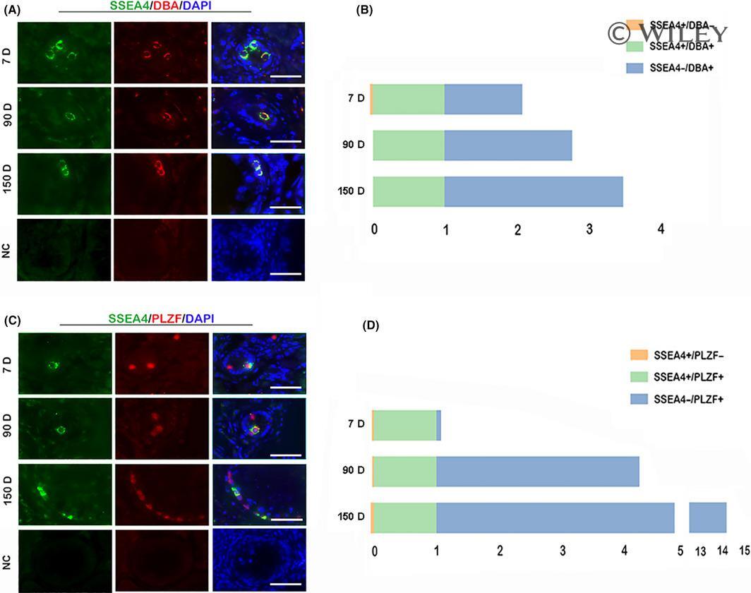
- 3 FIGURE SSEA4 was expressed in the undifferentiated spermatogonia. Co-immunofluorescence of SSEA4 and with the undifferentiated spermatogonial marker DBA (A) and PLZF (B) in testicular tissues from different ages. Relative proportion of SSEA4-positive cells co-localized with DBA-positive cells (C) and PLZF-positive cells (D). Data are normalized by the number of double-positive cells and are mean for six independent experiments. NC, Negative Control, staining without primary antibody. Scale bars = 50 mum
- Submitted by
- Invitrogen Antibodies (provider)
- Main image

- Experimental details
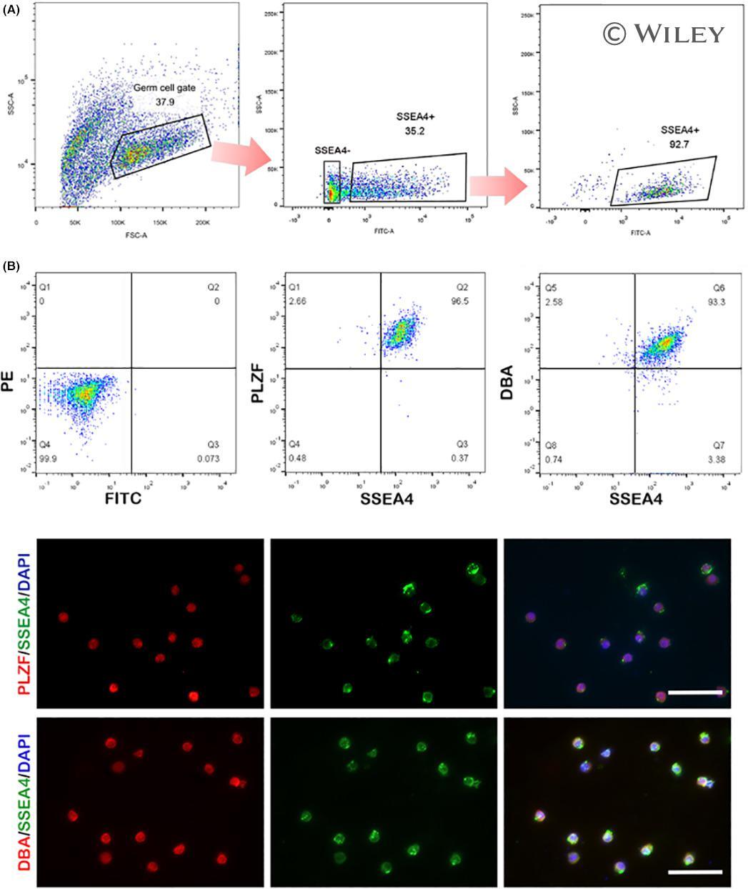
- 4 FIGURE Fluorescence-activated cell sorting (FACS) of the SSEA4-positive populations from the germ cells gate of 90-d-old porcine testes. (A) The FACS process of the SSEA4-positive cells. Left panel: The germ cell gate was evident on the light (forward and side) scatter dot plot from whole population. Middle panel: the SSEA4-positive and SSEA4-negative gate were sorted from germ cell gate. Right panel: the sorted SSEA4-positive cells were assayed by FACS. (B) Analysis of the percentage of PLZF (middle panel) and DBA-positive (right panel) cells in SEA4-positive fraction by FACS and immunocytochemical detection of PLZF/DBA in SEA4-positive fraction. Left panel in B: the negative control staining without the primary antibody. Scale bars = 50 mum