 Explore
 Validate
 Learn
Learn Western blot
 Immunocytochemistry
Antibody data
- Antibody Data
- Antigen structure
- References [0]
- Comments [0]
- Validations
- Western blot [1]
- Immunohistochemistry [7]
Submit
Validation data
Reference
Comment
Report error
- Product number
- LS-C359237 - Provider product page

- Provider
- LSBio
- Product name
- FOXP1 Antibody (clone JC12) LS-C359237
- Antibody type
- Monoclonal
- Description
- Protein G purified
- Reactivity
- Human, Mouse
- Host
- Mouse
- Isotype
- IgG
- Antibody clone number
- JC12
- Storage
- Short term: store at 4°C. Long term: aliquot and store at -20°C. Avoid freeze-thaw cycles.
No comments: Submit comment
Enhanced validation
- Submitted by
- LSBio (provider)
- Enhanced method
- Genetic validation
- Main image
- Experimental details
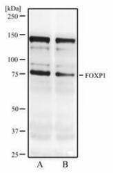
- Western Blot: FOXP1 Antibody (JC12) - Western blot analysis of resonicated MCF7 cell lysate (A) and MCF7 cell lysate (B) using FOXP1 antibody at 2 ug/ml.

Supportive validation
- Submitted by
- LSBio (provider)
- Enhanced method
- Genetic validation
- Main image

- Experimental details
- Immunohistochemistry-Paraffin: FOXP1 Antibody (JC12) - IHC analysis of formalin-fixed paraffin-embedded tissue section of malignant stromal tumor of the human small bowel using mouse monoclonal FOXP1 antibody (clone JC12) at 5 ug/ml concentration. The carcinoma cells developed an expected and specific strong nuclear with mild cytoplasmic immunopositivity for FOXP1 protein.

- Submitted by
- LSBio (provider)
- Enhanced method
- Genetic validation
- Main image

- Experimental details
- Immunohistochemistry-Paraffin: FOXP1 Antibody (JC12) - IHC analysis of formalin-fixed paraffin-embedded tissue section of human normal breast using FOXP1 antibody (clone JC12) at 5 ug/ml concentration. The breast ductal/acinar epithelial cells and the myoepithelial cells developed a strong nuclear along with moderate cytoplasmic immuno-positivity for FOXP1 protein.

- Submitted by
- LSBio (provider)
- Enhanced method
- Genetic validation
- Main image
- Experimental details
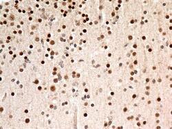
- Immunohistochemistry-Paraffin: FOXP1 Antibody (JC12) - IHC analysis of formalin-fixed paraffin-embedded tissue section of human normal brain using mouse monoclonal FOXP1 antibody (clone JC12) at 5 ug/ml concentration. The cells in the brain tissue depicted strong specific nuclear along with relatively weak cytoplasmic immunopositivity for FOXP1 protein.

- Submitted by
- LSBio (provider)
- Enhanced method
- Genetic validation
- Main image

- Experimental details
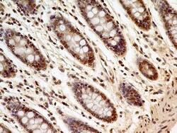
- Immunohistochemistry-Paraffin: FOXP1 Antibody (JC12) - IHC analysis of formalin-fixed paraffin-embedded tissue section of human normal colon using mouse monoclonal FOXP1 antibody (clone JC12) at 5 ug/ml concentration. Most of the cells depicted an expected strong nuclear with mild cytoplasmic staining.

- Submitted by
- LSBio (provider)
- Enhanced method
- Genetic validation
- Main image
- Experimental details
- Immunohistochemistry-Paraffin: FOXP1 Antibody (JC12) - IHC analysis of formalin-fixed paraffin-embedded tissue section of human normal colon using mouse monoclonal FOXP1 antibody (clone JC12) at 5 ug/ml concentration. Most of the cells depicted an expected strong nuclear with mild cytoplasmic staining.

- Submitted by
- LSBio (provider)
- Enhanced method
- Genetic validation
- Main image

- Experimental details
- Immunohistochemistry-Paraffin: FOXP1 Antibody (JC12) - IHC analysis of formalin-fixed paraffin-embedded tissue section of human normal brain using mouse monoclonal FOXP1 antibody (clone JC12) at 5 ug/ml concentration. The cells in the brain tissue depicted strong specific nuclear along with relatively weak cytoplasmic immunopositivity for FOXP1 protein.

- Submitted by
- LSBio (provider)
- Enhanced method
- Genetic validation
- Main image

- Experimental details
- Immunohistochemistry-Paraffin: FOXP1 Antibody (JC12) - IHC analysis of formalin-fixed paraffin-embedded tissue section of human normal colon using mouse monoclonal FOXP1 antibody (clone JC12) at 5 ug/ml concentration. Most of the cells depicted an expected strong nuclear with mild cytoplasmic staining.
