Explore
 Validate
 Learn
Learn Western blot
 Immunocytochemistry
Antibody data
- Antibody Data
- Antigen structure
- References [8]
- Comments [0]
- Validations
- Western blot [1]
- Immunohistochemistry [4]
- Flow cytometry [1]
Submit
Validation data
Reference
Comment
Report error
- Product number
- NB100-65125 - Provider product page

- Provider
- Novus Biologicals
- Proper citation
- Novus Cat#NB100-65125, RRID:AB_2106980
- Product name
- Mouse Monoclonal FoxP1 Antibody
- Antibody type
- Monoclonal
- Description
- Protein G purified. Does not recognize closely related molecules FOXP2, FOXP3 or FOXP4.
- Reactivity
- Human, Mouse
- Host
- Mouse
- Antigen sequence
Within the C-terminus- Isotype
- IgG
- Vial size
- 0.1 ml
- Concentration
- 1.0 mg/ml
- Storage
- Store at 4C short term. Aliquot and store at -20C long term. Avoid freeze-thaw cycles.
Submitted references The life and death of the germinal center.
p53 expression is a strong marker of inferior survival in de novo diffuse large B-cell lymphoma and may have enhanced negative effect with MYC coexpression: a single institutional clinicopathologic study.
Potentially oncogenic B-cell activation-induced smaller isoforms of FOXP1 are highly expressed in the activated B cell-like subtype of DLBCL.
FOXP3 is a homo-oligomer and a component of a supramolecular regulatory complex disabled in the human XLAAD/IPEX autoimmune disease.
Analysis of FOXP3 protein expression in human CD4(+)CD25(+) regulatory T cells at the single-cell level.
Expression of the FOXP1 transcription factor is strongly associated with inferior survival in patients with diffuse large B-cell lymphoma.
Expression of the forkhead transcription factor FOXP1 is associated with estrogen receptor alpha and improved survival in primary human breast carcinomas.
The FOXP1 winged helix transcription factor is a novel candidate tumor suppressor gene on chromosome 3p.
Gars E, Butzmann A, Ohgami R, Balakrishna JP, O'Malley DP
Annals of diagnostic pathology 2020 Feb;44:151421
Annals of diagnostic pathology 2020 Feb;44:151421
p53 expression is a strong marker of inferior survival in de novo diffuse large B-cell lymphoma and may have enhanced negative effect with MYC coexpression: a single institutional clinicopathologic study.
Xie Y, Bulbul MA, Ji L, Inouye CM, Groshen SG, Tulpule A, O'Malley DP, Wang E, Siddiqi IN
American journal of clinical pathology 2014 Apr;141(4):593-604
American journal of clinical pathology 2014 Apr;141(4):593-604
Potentially oncogenic B-cell activation-induced smaller isoforms of FOXP1 are highly expressed in the activated B cell-like subtype of DLBCL.
Brown PJ, Ashe SL, Leich E, Burek C, Barrans S, Fenton JA, Jack AS, Pulford K, Rosenwald A, Banham AH
Blood 2008 Mar 1;111(5):2816-24
Blood 2008 Mar 1;111(5):2816-24
FOXP3 is a homo-oligomer and a component of a supramolecular regulatory complex disabled in the human XLAAD/IPEX autoimmune disease.
Li B, Samanta A, Song X, Iacono KT, Brennan P, Chatila TA, Roncador G, Banham AH, Riley JL, Wang Q, Shen Y, Saouaf SJ, Greene MI
International immunology 2007 Jul;19(7):825-35
International immunology 2007 Jul;19(7):825-35
Analysis of FOXP3 protein expression in human CD4(+)CD25(+) regulatory T cells at the single-cell level.
Loddenkemper C, Maul J, Berg E, Stein H, Zeitz M, Duchmann R
European journal of immunology 2006 Jan;36(1):245; author reply 246
European journal of immunology 2006 Jan;36(1):245; author reply 246
Expression of the FOXP1 transcription factor is strongly associated with inferior survival in patients with diffuse large B-cell lymphoma.
Banham AH, Connors JM, Brown PJ, Cordell JL, Ott G, Sreenivasan G, Farinha P, Horsman DE, Gascoyne RD
Clinical cancer research : an official journal of the American Association for Cancer Research 2005 Feb 1;11(3):1065-72
Clinical cancer research : an official journal of the American Association for Cancer Research 2005 Feb 1;11(3):1065-72
Expression of the forkhead transcription factor FOXP1 is associated with estrogen receptor alpha and improved survival in primary human breast carcinomas.
Fox SB, Brown P, Han C, Ashe S, Leek RD, Harris AL, Banham AH
Clinical cancer research : an official journal of the American Association for Cancer Research 2004 May 15;10(10):3521-7
Clinical cancer research : an official journal of the American Association for Cancer Research 2004 May 15;10(10):3521-7
The FOXP1 winged helix transcription factor is a novel candidate tumor suppressor gene on chromosome 3p.
Banham AH, Beasley N, Campo E, Fernandez PL, Fidler C, Gatter K, Jones M, Mason DY, Prime JE, Trougouboff P, Wood K, Cordell JL
Cancer research 2001 Dec 15;61(24):8820-9
Cancer research 2001 Dec 15;61(24):8820-9
No comments: Submit comment
Supportive validation
- Submitted by
- Novus Biologicals (provider)
- Main image
- Experimental details
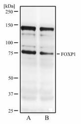
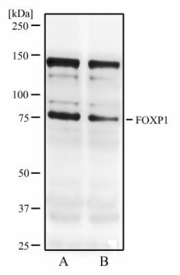
- Western Blot: FOXP1 Antibody (JC12) [NB100-65125] - Western blot analysis of resonicated MCF7 cell lysate (A) and MCF7 cell lysate (B) using FOXP1 antibody at 2 ug/ml.
Supportive validation
- Submitted by
- Novus Biologicals (provider)
- Main image
- Experimental details
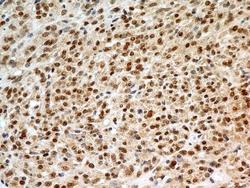
- Immunohistochemistry-Paraffin: FOXP1 Antibody (JC12) [NB100-65125] - IHC analysis of formalin-fixed paraffin-embedded tissue section of malignant stromal tumor of the human small bowel using mouse monoclonal FOXP1 antibody (clone JC12) at 5 ug/ml concentration. The carcinoma cells developed an expected and specific strong nuclear with mild cytoplasmic immunopositivity for FOXP1 protein.
- Submitted by
- Novus Biologicals (provider)
- Main image
- Experimental details
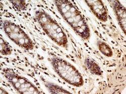
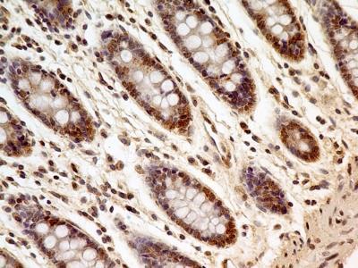
- Immunohistochemistry-Paraffin: FOXP1 Antibody (JC12) [NB100-65125] - IHC analysis of formalin-fixed paraffin-embedded tissue section of human normal colon using mouse monoclonal FOXP1 antibody (clone JC12) at 5 ug/ml concentration. Most of the cells depicted an expected strong nuclear with mild cytoplasmic staining.
- Submitted by
- Novus Biologicals (provider)
- Main image

- Experimental details
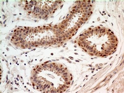
- Immunohistochemistry-Paraffin: FOXP1 Antibody (JC12) [NB100-65125] - IHC analysis of formalin-fixed paraffin-embedded tissue section of human normal breast using FOXP1 antibody (clone JC12) at 5 ug/ml concentration. The breast ductal/acinar epithelial cells and the myoepithelial cells developed a strong nuclear along with moderate cytoplasmic immuno-positivity for FOXP1 protein.
- Submitted by
- Novus Biologicals (provider)
- Main image
- Experimental details
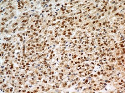
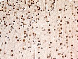
- Immunohistochemistry-Paraffin: FOXP1 Antibody (JC12) [NB100-65125] - IHC analysis of formalin-fixed paraffin-embedded tissue section of human normal brain using mouse monoclonal FOXP1 antibody (clone JC12) at 5 ug/ml concentration. The cells in the brain tissue depicted strong specific nuclear along with relatively weak cytoplasmic immunopositivity for FOXP1 protein.

Supportive validation
- Submitted by
- Novus Biologicals (provider)
- Main image

- Experimental details
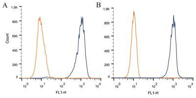
- Flow Cytometry: FOXP1 Antibody (JC12) [NB100-65125] - Intracellular flow cytometric staining of 1 x 10^6 CHO (A) and HeLa (B) cells using FOXP1 antibody (dark blue). Isotype control shown in orange. An antibody concentration of 1 ug/1x10^6 cells was used.