Explore
 Validate
 Learn
Learn Western blot
Antibody data
- Antibody Data
- Antigen structure
- References [3]
- Comments [0]
- Validations
- Western blot [4]
- Immunocytochemistry [5]
- Immunohistochemistry [2]
- Other assay [2]
Submit
Validation data
Reference
Comment
Report error
- Product number
- PA5-88084 - Provider product page

- Provider
- Invitrogen Antibodies
- Product name
- Nrf2 Polyclonal Antibody
- Antibody type
- Polyclonal
- Antigen
- Synthetic peptide
- Reactivity
- Human, Mouse, Rat
- Host
- Rabbit
- Isotype
- IgG
- Vial size
- 100 µL
- Concentration
- 0.92 mg/mL
- Storage
- -20° C, Avoid Freeze/Thaw Cycles
Submitted references Short-term exposure to JUUL electronic cigarettes can worsen ischemic stroke outcome.
Lysosomal nitric oxide determines transition from autophagy to ferroptosis after exposure to plasma-activated Ringer's lactate.
Impact of chronic smoking on traumatic brain microvascular injury: An in vitro study.
Sifat AE, Archie SR, Nozohouri S, Villalba H, Zhang Y, Sharma S, Ghanwatkar Y, Vaidya B, Mara D, Cucullo L, Abbruscato TJ
Fluids and barriers of the CNS 2022 Sep 9;19(1):74
Fluids and barriers of the CNS 2022 Sep 9;19(1):74
Lysosomal nitric oxide determines transition from autophagy to ferroptosis after exposure to plasma-activated Ringer's lactate.
Jiang L, Zheng H, Lyu Q, Hayashi S, Sato K, Sekido Y, Nakamura K, Tanaka H, Ishikawa K, Kajiyama H, Mizuno M, Hori M, Toyokuni S
Redox biology 2021 Jul;43:101989
Redox biology 2021 Jul;43:101989
Impact of chronic smoking on traumatic brain microvascular injury: An in vitro study.
Sivandzade F, Alqahtani F, Cucullo L
Journal of cellular and molecular medicine 2021 Aug;25(15):7122-7134
Journal of cellular and molecular medicine 2021 Aug;25(15):7122-7134
No comments: Submit comment
Supportive validation
- Submitted by
- Invitrogen Antibodies (provider)
- Main image

- Experimental details
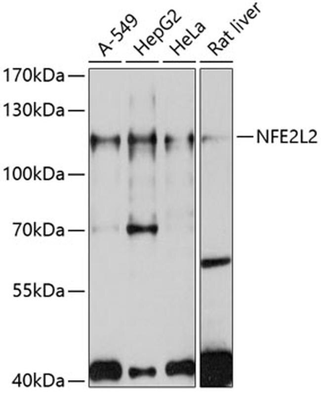
- Western blot analysis of extracts of various cell lines, using NFE2L2 Polyclonal antibody (Product # PA5-88084) at 1:1000 dilution. Secondary antibody: HRP Goat Anti-Rabbit IgG (H+L) at 1:10000 dilution. Lysates/proteins: 25ug per lane. Blocking buffer: 3% nonfat dry milk in TBST. Exposure time: 10s.
- Submitted by
- Invitrogen Antibodies (provider)
- Main image
- Experimental details
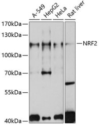
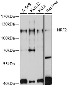
- Western Blot analysis of Nrf2 in extracts of various cell lines using Nrf2 Polyclonal Antibody (Product # PA5-88084) at a dilution of 1:1000. A HRP Goat Anti-Rabbit IgG (H+L) secondary antibody was used at a dilution of 1:10,000. Lysates/proteins: 25 µg per lane. Blocking buffer: 3% nonfat dry milk in TBST.
- Submitted by
- Invitrogen Antibodies (provider)
- Main image

- Experimental details
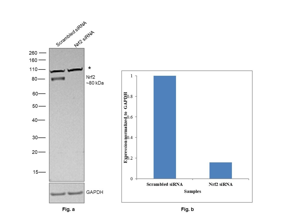
- Knockdown of Nuclear factor erythroid 2-related factor 2 was achieved by transfecting U-2 OS cells treated with MG-132 (10 micromolar for 5h) with Nuclear factor erythroid 2-related factor 2 specific siRNAs (Silencer® select Product # s9493, s9492). Western blot analysis (Fig. a) was performed using whole-cell extracts from the Nuclear factor erythroid 2-related factor 2 knockdown cells (lane 2), and non-targeting scrambled siRNA transfected cells (lane 1). The blot was probed with Nrf2 Polyclonal Antibody (Product # PA5-88084, 1:1000 dilution ) and Goat anti-Rabbit IgG (H+L) Superclonal™ Recombinant Secondary Antibody, HRP (Product # A27036, 1:4000 dilution). Densitometric analysis of this western blot is shown in histogram (Fig. b). Decrease in signal upon siRNA mediated knockdown confirms that antibody is specific to Nuclear factor erythroid 2-related factor 2. An uncharacterized band (*) was observed at ~110 kDa that was not downregulated upon siRNA transfection.
- Submitted by
- Invitrogen Antibodies (provider)
- Main image
- Experimental details
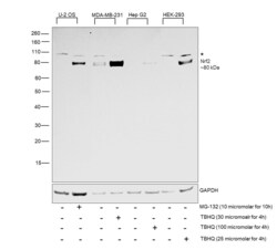
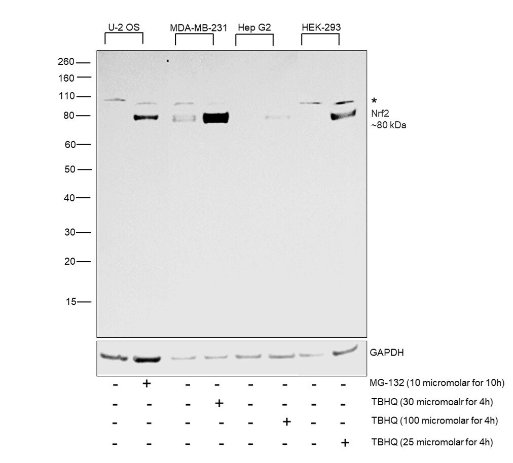
- Western blot was performed using Anti-Nrf2 Polyclonal Antibody (Product # PA5-88084) and a 80kDa band corresponding to Nuclear factor erythroid 2-related factor 2 was observed to be upregulated upon MG-132 and TBHQ treatment across various cell lines. Whole-cell extracts (30 µg lysate) of U-2 OS (Lane 1), U-2 OS treated with MG-132 (10 micromolar for 10h) (Lane 2), MDA-MB-231 (Lane 3), MDA-MB-231 treated with TBHQ (30 micromolar for 4h) (Lane 4), Hep G2 (Lane 5), Hep G2 treated with TBHQ (100 micromolar for 4h) (Lane 6), HEK-293 (Lane 7), HEK-293 treated with TBHQ (25 micromolar for 4h) (Lane 8) were electrophoresed using NuPAGE™ 4-12% Bis-Tris Protein Gel (Product # NP0322BOX). Resolved proteins were then transferred onto a Nitrocellulose membrane (Product # IB23001) by iBlot® 2 Dry Blotting System (Product # IB21001). The blot was probed with the primary antibody (1:1000 dilution) and detected by chemiluminescence with Goat anti-Rabbit IgG (H+L) Superclonal™ Recombinant Secondary Antibody, HRP (Product # A27036, 1:4000 dilution) using the iBright FL 1000 (Product # A32752). Chemiluminescent detection was performed using SuperSignal™ West Dura Extended Duration Substrate (Product # 34076).
Supportive validation
- Submitted by
- Invitrogen Antibodies (provider)
- Main image
- Experimental details
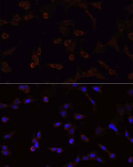
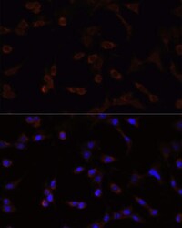
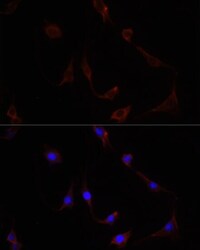
- Immunocytochemistry-Immunofluorescence analysis of Nrf2 was performed in NIH/3T3 cells using Nrf2 Polyclonal Antibody (Product # PA5-88084).
- Submitted by
- Invitrogen Antibodies (provider)
- Main image
- Experimental details
- Immunocytochemistry-Immunofluorescence analysis of Nrf2 was performed in PC12 cells using Nrf2 Polyclonal Antibody (Product # PA5-88084).
- Submitted by
- Invitrogen Antibodies (provider)
- Main image

- Experimental details
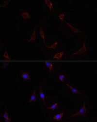
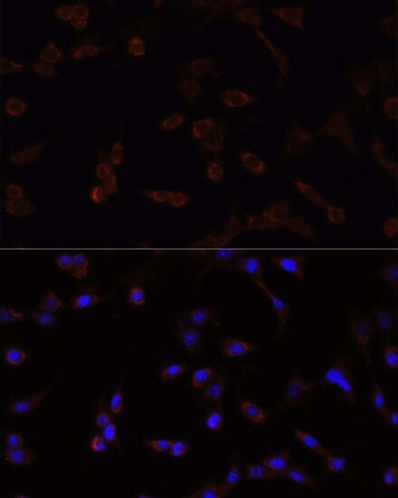
- Immunocytochemistry-Immunofluorescence analysis of Nrf2 was performed in HeLa cells using Nrf2 Polyclonal Antibody (Product # PA5-88084).
- Submitted by
- Invitrogen Antibodies (provider)
- Main image
- Experimental details
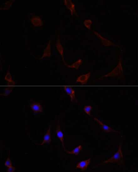
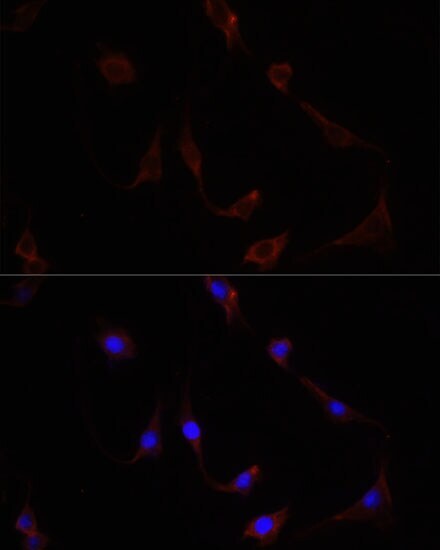
- Immunocytochemistry-Immunofluorescence analysis of Nrf2 was performed in NIH/3T3 cells using Nrf2 Polyclonal Antibody (Product # PA5-88084).
- Submitted by
- Invitrogen Antibodies (provider)
- Main image
- Experimental details
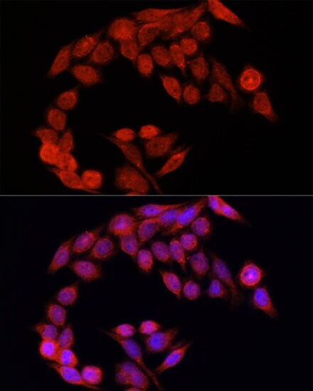
- Immunocytochemistry-Immunofluorescence analysis of Nrf2 was performed in PC12 cells using Nrf2 Polyclonal Antibody (Product # PA5-88084).
Supportive validation
- Submitted by
- Invitrogen Antibodies (provider)
- Main image
- Experimental details
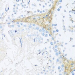
- Immunohistochemistry analysis of Nrf2 in paraffin-embedded mouse leydig cells using Nrf2 Polyclonal Antibody (Product # PA5-88084) at a dilution of 1:200.

- Submitted by
- Invitrogen Antibodies (provider)
- Main image

- Experimental details
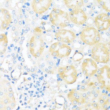
- Immunohistochemistry analysis of Nrf2 in paraffin-embedded mouse kidney using Nrf2 Polyclonal Antibody (Product # PA5-88084) at a dilution of 1:200.
Supportive validation
- Submitted by
- Invitrogen Antibodies (provider)
- Main image

- Experimental details
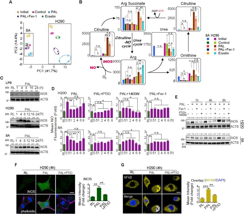
- Fig. 2 PAL effects not only through exogenous but also through endogenous nitric oxide ( . NO) on MM cells. (A) Principal components analysis (PCA) of metabolome reveals a separation of main source of variance among initial (cells cultured in complete RPMI1640 medium), control (cells cultured in RL solution), PAL treatment, PAL + Fer-1 and erastin-treatment (N = 3; H290 sarcomatoid MM; 8A, epithelioid MM). (B) Mapping of metabolite levels in citrulline- . NO cycle and urea cycle. Comparison of relative metabolite levels in cells cultivated in complete medium (initial), RL solution (control), PAL, PAL + Fer-1 or erastin. Relative metabolite levels are shown as bar plots. Characteristic reactions of citrulline- . NO cycle and urea cycle are included. (C) Specific PAL-mediated iNOS induction in MM cells. Human mesothelial cells (LP9) and MM cells (H290 and 8A) were treated with PAL (15 min) and then incubated in medium for 0-24 h/37 degC, followed by Western blot analysis. The numbers above the bands indicate relative values by quantification through the entire figures. (D) Biphasic . NO increase. MM cells were treated with or without PAL for 15 min. The cells were then incubated with or without caboxy-PTIO, 1400 W or Fer-1 for an additional 0.5-8 h/37 degC, and . NO was detected by flow cytometry using the DAF-FM DA probe. (E) Association of . NO and iNOS expression. MM cells were treated with PAL (15 min) and then incubated in medium for 0-8 h/37 degC with or without Fer-1/a c
- Submitted by
- Invitrogen Antibodies (provider)
- Main image
- Experimental details
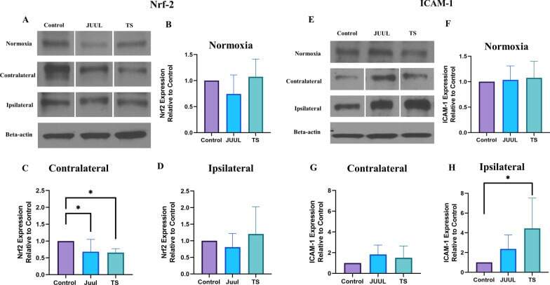
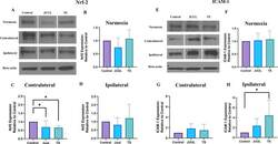
- Brain Nrf2 and ICAM-1 expression in mice after 14 days of JUUL/TS exposure with or without MCAO. A Western blot images of Nrf2 expression in normoxic brain and contralateral & ipsilateral brain hemispheres 24 h after MCAO. B - D Quantification of brain Nrf2 expression normalized to beta-actin and expressed as relative to control (1.0) in normoxic, contralateral, and ipsilateral brain regions. E Western blot images of ICAM-1 expression in normoxic brain and contralateral & ipsilateral brain hemispheres 24 h after MCAO. F - H Quantification of brain ICAM-1 expression normalized to beta-actin and expressed as relative to control (1.0) in normoxic, contralateral, and ipsilateral brain regions. *P < 0.05; n = 9 for each group (B), n = 7, 7, and 6 for control, JUUL, and TS respectively ( C , D ), n = 9 for each group ( F ), n = 6, 5, and 5 for control, JUUL, and TS respectively ( G , H ). Cropped images from blots have been used in some cases to improve the clarity and conciseness of the presentation. The cropped images are delineated with white space; full-length blots developed by X-ray films are presented in Additional file 1 : Fig. S1