Antibody data
- Antibody Data
- Antigen structure
- References [13]
- Comments [0]
- Validations
- Western blot [2]
- Immunocytochemistry [1]
- Immunohistochemistry [3]
- Other assay [6]
Submit
Validation data
Reference
Comment
Report error
- Product number
- PA1-520 - Provider product page

- Provider
- Invitrogen Antibodies
- Product name
- CLOCK Polyclonal Antibody
- Antibody type
- Polyclonal
- Antigen
- Synthetic peptide
- Description
- PA1-520 detects CLOCK from human, mouse, rat and hamster tissues. PA1-520 has been successfully used in Western blot, ICC/IF and immunohistochemistry procedures. By Western blot, this antibody detects an ~100 kDa protein representing CLOCK from rat brain homogenate. Immunohistochemical staining of CLOCK in hamster brain with PA1-520 results in the staining of the superchiasmatic nucleus. PA1-520 immunizing peptide corresponds to amino acid residues 11-30 of mouse CLOCK. This sequence is completely conserved between mouse and human CLOCK. PA1-520 immunizing peptide (Cat. # PEP-072) is available for use in neutralization and control experiments.
- Reactivity
- Human, Mouse, Rat, Hamster
- Host
- Rabbit
- Isotype
- IgG
- Vial size
- 200 µL
- Concentration
- 1 mg/mL
- Storage
- -20° C, Avoid Freeze/Thaw Cycles
Submitted references Prolonged Administration of Melatonin Ameliorates Liver Phenotypes in Cholestatic Murine Model.
Circadian Oscillations Persist in Cervical and Esophageal Cancer Cells Displaying Decreased Expression of Tumor-Suppressing Circadian Clock Genes.
Melatonin modulates daytime-dependent synaptic plasticity and learning efficiency.
Comparative cell cycle transcriptomics reveals synchronization of developmental transcription factor networks in cancer cells.
Circadian networks in human embryonic stem cell-derived cardiomyocytes.
Time-caloric restriction inhibits the neoplastic transformation of cirrhotic liver in rats treated with diethylnitrosamine.
Diurnal oscillation of amygdala clock gene expression and loss of synchrony in a mouse model of depression.
p53 regulates Period2 expression and the circadian clock.
Circadian rhythms regulate amelogenesis.
Expression of clock proteins in developing tooth.
Day-night expression patterns of clock genes in the human pineal gland.
The E2F-regulated gene Chk1 is highly expressed in triple-negative estrogen receptor /progesterone receptor /HER-2 breast carcinomas.
Non-cyclic and developmental stage-specific expression of circadian clock proteins during murine spermatogenesis.
Ceci L, Chen L, Baiocchi L, Wu N, Kennedy L, Carpino G, Kyritsi K, Zhou T, Owen T, Kundu D, Sybenga A, Isidan A, Ekser B, Franchitto A, Onori P, Gaudio E, Mancinelli R, Francis H, Alpini G, Glaser S
Cellular and molecular gastroenterology and hepatology 2022;14(4):877-904
Cellular and molecular gastroenterology and hepatology 2022;14(4):877-904
Circadian Oscillations Persist in Cervical and Esophageal Cancer Cells Displaying Decreased Expression of Tumor-Suppressing Circadian Clock Genes.
van der Watt PJ, Roden LC, Davis KT, Parker MI, Leaner VD
Molecular cancer research : MCR 2020 Sep;18(9):1340-1353
Molecular cancer research : MCR 2020 Sep;18(9):1340-1353
Melatonin modulates daytime-dependent synaptic plasticity and learning efficiency.
Jilg A, Bechstein P, Saade A, Dick M, Li TX, Tosini G, Rami A, Zemmar A, Stehle JH
Journal of pineal research 2019 Apr;66(3):e12553
Journal of pineal research 2019 Apr;66(3):e12553
Comparative cell cycle transcriptomics reveals synchronization of developmental transcription factor networks in cancer cells.
Boström J, Sramkova Z, Salašová A, Johard H, Mahdessian D, Fedr R, Marks C, Medalová J, Souček K, Lundberg E, Linnarsson S, Bryja V, Sekyrova P, Altun M, Andäng M
PloS one 2017;12(12):e0188772
PloS one 2017;12(12):e0188772
Circadian networks in human embryonic stem cell-derived cardiomyocytes.
Dierickx P, Vermunt MW, Muraro MJ, Creyghton MP, Doevendans PA, van Oudenaarden A, Geijsen N, Van Laake LW
EMBO reports 2017 Jul;18(7):1199-1212
EMBO reports 2017 Jul;18(7):1199-1212
Time-caloric restriction inhibits the neoplastic transformation of cirrhotic liver in rats treated with diethylnitrosamine.
Molina-Aguilar C, Guerrero-Carrillo MJ, Espinosa-Aguirre JJ, Olguin-Reyes S, Castro-Belio T, Vázquez-Martínez O, Rivera-Zavala JB, Díaz-Muñoz M
Carcinogenesis 2017 Aug 1;38(8):847-858
Carcinogenesis 2017 Aug 1;38(8):847-858
Diurnal oscillation of amygdala clock gene expression and loss of synchrony in a mouse model of depression.
Savalli G, Diao W, Schulz S, Todtova K, Pollak DD
The international journal of neuropsychopharmacology 2014 Dec 11;18(5)
The international journal of neuropsychopharmacology 2014 Dec 11;18(5)
p53 regulates Period2 expression and the circadian clock.
Miki T, Matsumoto T, Zhao Z, Lee CC
Nature communications 2013;4:2444
Nature communications 2013;4:2444
Circadian rhythms regulate amelogenesis.
Zheng L, Seon YJ, Mourão MA, Schnell S, Kim D, Harada H, Papagerakis S, Papagerakis P
Bone 2013 Jul;55(1):158-65
Bone 2013 Jul;55(1):158-65
Expression of clock proteins in developing tooth.
Zheng L, Papagerakis S, Schnell SD, Hoogerwerf WA, Papagerakis P
Gene expression patterns : GEP 2011 Mar-Apr;11(3-4):202-6
Gene expression patterns : GEP 2011 Mar-Apr;11(3-4):202-6
Day-night expression patterns of clock genes in the human pineal gland.
Ackermann K, Dehghani F, Bux R, Kauert G, Stehle JH
Journal of pineal research 2007 Sep;43(2):185-94
Journal of pineal research 2007 Sep;43(2):185-94
The E2F-regulated gene Chk1 is highly expressed in triple-negative estrogen receptor /progesterone receptor /HER-2 breast carcinomas.
Verlinden L, Vanden Bempt I, Eelen G, Drijkoningen M, Verlinden I, Marchal K, De Wolf-Peeters C, Christiaens MR, Michiels L, Bouillon R, Verstuyf A
Cancer research 2007 Jul 15;67(14):6574-81
Cancer research 2007 Jul 15;67(14):6574-81
Non-cyclic and developmental stage-specific expression of circadian clock proteins during murine spermatogenesis.
Alvarez JD, Chen D, Storer E, Sehgal A
Biology of reproduction 2003 Jul;69(1):81-91
Biology of reproduction 2003 Jul;69(1):81-91
No comments: Submit comment
Supportive validation
- Submitted by
- Invitrogen Antibodies (provider)
- Main image
- Experimental details
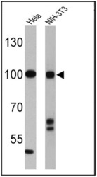
- Western blot analysis of CLOCK was performed by loading 25 µg of Hela (lane 1) and NIH-3T3 (lane 2) cell lysates onto an SDS polyacrylamide gel. Proteins were transferred to a PVDF membrane and blocked at 4ºC overnight. The membrane was probed with a CLOCK polyclonal antibody (Product # PA1-520) at a dilution of 1:4000 overnight at 4°C, washed in TBST, and probed with an HRP-conjugated secondary antibody for 1 hr at room temperature in the dark. Chemiluminescent detection was performed using Pierce ECL Plus Western Blotting Substrate (Product # 32132). Results show a band at ~100 kDa.
- Submitted by
- Invitrogen Antibodies (provider)
- Main image
- Experimental details
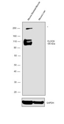
- Western blot was performed using Anti-CLOCK Polyclonal Antibody (Product # PA1-520) and a ~95 kDa band corresponding to CLOCK was observed in Mouse Skeletal Muscle and not in Mouse Liver which is reported to be negative. An uncharacterized band (*) at ~260 kDa was observed in Mouse Skeletal Muscle. Tissue extracts (30ug) of Mouse Skeletal Muscle (Lane 1), Mouse Liver (Lane 2) were electrophoresed using Novex® NuPAGE® 4-12 % Bis-Tris gel (Product # NP0322BOX). Resolved proteins were then transferred onto a nitrocellulose membrane (Product # IB23001) by iBlot® 2 Dry Blotting System (Product # IB21001). The blot was probed with the primary antibody (1:2000 dilution) and detected by chemiluminescence with Goat anti-Rabbit IgG (H+L) Superclonal™ Recombinant Secondary Antibody, HRP (Product # A27036, 1:4000 dilution) using the iBright FL 1000 (Product # A32752). Chemiluminescent detection was performed using Novex® ECL Chemiluminescent Substrate Reagent Kit (Product # WP20005).
Supportive validation
- Submitted by
- Invitrogen Antibodies (provider)
- Main image
- Experimental details
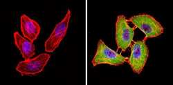
- Immunofluorescent analysis of CLOCK (green) showing staining in the nucleus and cytoplasm of U251 cells (right) compared to a negative control without primary antibody (left). Formalin-fixed cells were permeabilized with 0.1% Triton X-100 in TBS for 5-10 minutes and blocked with 3% BSA-PBS for 30 minutes at room temperature. Cells were probed with a CLOCK polyclonal antibody (Product # PA1-520) in 3% BSA-PBS at a dilution of 1:100 and incubated overnight at 4 ºC in a humidified chamber. Cells were washed with PBST and incubated with a DyLight-conjugated secondary antibody in PBS at room temperature in the dark. F-actin (red) was stained with a fluorescent red phalloidin and nuclei (blue) were stained with Hoechst or DAPI. Images were taken at a magnification of 60x.
Supportive validation
- Submitted by
- Invitrogen Antibodies (provider)
- Main image
- Experimental details
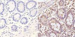
- Immunohistochemistry analysis of CLOCK showing positive staining in the nucleus and cytoplasm of paraffin-treated Human colon tissue (right) compared with a negative control in the absence of primary antibody (left). To expose target proteins, antigen retrieval method was performed using 10mM sodium citrate (pH 6.0) microwaved for 8-15 min. Following antigen retrieval, tissues were blocked in 3% H2O2-methanol for 15 min at room temperature, washed with ddH2O and PBS, and then probed with a CLOCK polyclonal antibody (Product # PA1-520) diluted by 3% BSA-PBS at a dilution of 1:200 overnight at 4°C in a humidified chamber. Tissues were washed extensively PBST and detection was performed using an HRP-conjugated secondary antibody followed by colorimetric detection using a DAB kit. Tissues were counterstained with hematoxylin and dehydrated with ethanol and xylene to prep for mounting.
- Submitted by
- Invitrogen Antibodies (provider)
- Main image
- Experimental details
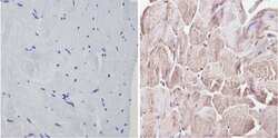
- Immunohistochemistry analysis of CLOCK showing positive staining in the nucleus and cytoplasm of paraffin-treated Human skeletal muscle (right) compared with a negative control in the absence of primary antibody (left). To expose target proteins, antigen retrieval method was performed using 10mM sodium citrate (pH 6.0) microwaved for 8-15 min. Following antigen retrieval, tissues were blocked in 3% H2O2-methanol for 15 min at room temperature, washed with ddH2O and PBS, and then probed with a CLOCK polyclonal antibody (Product # PA1-520) diluted by 3% BSA-PBS at a dilution of 1:200 overnight at 4°C in a humidified chamber. Tissues were washed extensively PBST and detection was performed using an HRP-conjugated secondary antibody followed by colorimetric detection using a DAB kit. Tissues were counterstained with hematoxylin and dehydrated with ethanol and xylene to prep for mounting.
- Submitted by
- Invitrogen Antibodies (provider)
- Main image
- Experimental details
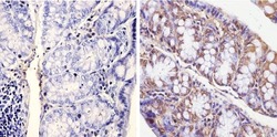
- Immunohistochemistry analysis of CLOCK showing positive staining in the nucleus and cytoplasm of paraffin-treated Mouse colon tissue (right) compared with a negative control in the absence of primary antibody (left). To expose target proteins, antigen retrieval method was performed using 10mM sodium citrate (pH 6.0) microwaved for 8-15 min. Following antigen retrieval, tissues were blocked in 3% H2O2-methanol for 15 min at room temperature, washed with ddH2O and PBS, and then probed with a CLOCK polyclonal antibody (Product # PA1-520) diluted by 3% BSA-PBS at a dilution of 1:200 overnight at 4°C in a humidified chamber. Tissues were washed extensively PBST and detection was performed using an HRP-conjugated secondary antibody followed by colorimetric detection using a DAB kit. Tissues were counterstained with hematoxylin and dehydrated with ethanol and xylene to prep for mounting.
Supportive validation
- Submitted by
- Invitrogen Antibodies (provider)
- Main image
- Experimental details
- NULL
- Submitted by
- Invitrogen Antibodies (provider)
- Main image
- Experimental details
- NULL
- Submitted by
- Invitrogen Antibodies (provider)
- Main image
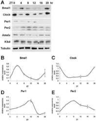
- Experimental details
- NULL
- Submitted by
- Invitrogen Antibodies (provider)
- Main image
- Experimental details
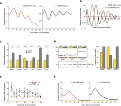
- Figure 1 Non-oscillatory expression of clock genes in human ES cells Raw lentiviral promoter-based luciferase reporter bioluminescence in U2OS cells after dexamethasone synchronization. Bioluminescence was measured with a LumiCycle32. Values are relative to T0. Detrended bioluminescent signals measured in (A). BMAL1, PER2 , CRY1 , CRY2 , CLOCK , and NR1D1 expression levels in human ES cells compared to U2OS cells as determined by qRT-PCR. Expression levels were normalized to PPIA and compared between cell types using an unpaired two-tailed Student's t -test (ns: not significant, * P < 0.05, ** P < 0.005, and *** P < 0.0005). Data are represented as mean +- s.e.m. of three independent replicates. Western blot for BMAL1, CRY1, and CLOCK. Protein levels were quantified and normalized to beta-ACTIN. qRT-PCR analysis of BMAL1 and PER2 expression over 48 h at a 4-h interval in human ES cells. Circadian oscillations were analyzed using the RAIN algorithm, and the significance of rhythmicity across 48 h is indicated (ns: not significant). Data are represented as mean +- s.e.m. of three independent replicates. Bmal1-dLuc and Per2-dLuc values in synchronized human ES cells across 76 h measured by LumiCycle32. Representative tracks are shown. Values are relative to T0.
- Submitted by
- Invitrogen Antibodies (provider)
- Main image
- Experimental details
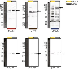
- Figure EV1 Protein levels of clock genes in human ES cells versus U2OS cells Original Western blots for BMAL1, CRY1, and CLOCK protein.
- Submitted by
- Invitrogen Antibodies (provider)
- Main image
- Experimental details
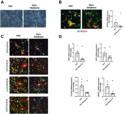
- Melatonin influences the phenotype of isolated P92-PSC. ( A ) Morphologic shapes of human isolated PSC cell line. Scale bars : 20 mum. ( B ) By immunofluorescence and qPCR, the MT1 and ( C and D ) clock genes (PER1, CRY1, CLOCK, and BMAL1) are decreased in isolated PSC cell lines treated with melatonin compared with vehicle-treated PSC cells. Original magnification, 20x; scale bars : 20 mum. Data are means +- SEM of 2 evaluations from 3 cumulative preparations of in vitro experiments. Each dot represents 1 value in data set. * P < .05 vs PSC.
 Explore
 Validate
 Learn
Learn Western blot