 Explore
 Validate
 Learn
Learn Western blot
 Immunocytochemistry
 Immunohistochemistry
Antibody data
- Antibody Data
- Antigen structure
- References [7]
- Comments [0]
- Validations
- Immunocytochemistry [4]
- Chromatin Immunoprecipitation [2]
Submit
Validation data
Reference
Comment
Report error
- Product number
- 14-9765-80 - Provider product page

- Provider
- Invitrogen Antibodies
- Product name
- SOX9 Monoclonal Antibody (GMPR9), eBioscience™
- Antibody type
- Monoclonal
- Antigen
- Other
- Description
- Description: This GMPR9 monoclonal antibody reacts with human and mouse Sox9, a member of the Sry HMG-box (Sox) gene family of transcription factors. Like Sry, Sox9 contains a high mobility group (HMG-box) DNA-binding domain. The Sox9 DNA-binding domain recognizes the sequence AGAACAATGG, and when bound to co-factors, functions to bend the DNA. This structural change is thought to be important for chromatin remodeling and the initiation of more broad transcriptional regulation of multiple genes. Sox9 is expressed in multiple tissue types during development and its activation of downstream targets is determined by interaction with tissue-specific co-factors. During development, Sox9 expression is important for cartilage formation (mesenchymal to chondrocyte), sex determination (testes formation), neural crest formation and retinal development. Sox9 plays a role in the differentiation of multiple cell types including glial, hair, cardiac, melanocytes, pancreatic, prostate, and intestinal epithelium. Sox9 regulates expression of the following family member genes sox5, sox6, and sox10, in addition to collagen type 2 alpha, collagen 9A1, CEACAM1, and snail2. Sox9 dysregulation is involved in craniofacial defects, cleft palate, sex reversal, and some types of cancer (melanoma, lung adenocarcinoma, and breast cancer). Sox9 has also been shown to be expressed in adult stem cells in the liver, pancreas and intestine. Applications Reported: This GMPR9 antibody has been reported for use in western blotting, immunohistochemical staining of formalin-fixed paraffin embedded tissue sections, microscopy, and immunocytochemistry. Applications Tested: This GMPR9 antibody has been tested by immunohistochemistry of formalin fixed paraffin embedded human tissue using high or low pH antigen retrieval and can be used at less than or equal to 1 µg/mL. This GMPR9 antibody has been tested by immunocytochemistry of fixed and permeabilized human cells and can be used at less than or equal to 1 µg/mL. It is recommended that the antibody be carefully titrated for optimal performance in the assay of interest. Purity: Greater than 90%, as determined by SDS-PAGE. Aggregation: Less than 10%, as determined by HPLC. Filtration: 0.2 µm post-manufacturing filtered.
- Reactivity
- Human, Mouse, Rat
- Host
- Mouse
- Isotype
- IgG
- Antibody clone number
- GMPR9
- Vial size
- 25 μg
- Concentration
- 0.5 mg/mL
- Storage
- 4°C
Submitted references SIX2 Regulates Human β Cell Differentiation from Stem Cells and Functional Maturation In Vitro.
Disruption of a hedgehog-foxf1-rspo2 signaling axis leads to tracheomalacia and a loss of sox9+ tracheal chondrocytes.
The sox family of transcription factors: versatile regulators of stem and progenitor cell fate.
Sox9 and NFIA coordinate a transcriptional regulatory cascade during the initiation of gliogenesis.
Sox9 marks adult organ progenitors.
Sox proteins in melanocyte development and melanoma.
Ectopic SOX9 mediates extracellular matrix deposition characteristic of organ fibrosis.
Velazco-Cruz L, Goedegebuure MM, Maxwell KG, Augsornworawat P, Hogrebe NJ, Millman JR
Cell reports 2020 May 26;31(8):107687
Cell reports 2020 May 26;31(8):107687
Disruption of a hedgehog-foxf1-rspo2 signaling axis leads to tracheomalacia and a loss of sox9+ tracheal chondrocytes.
Nasr T, Holderbaum AM, Chaturvedi P, Agarwal K, Kinney JL, Daniels K, Trisno SL, Ustiyan V, Shannon JM, Wells JM, Sinner D, Kalinichenko VV, Zorn AM
Disease models & mechanisms 2020 Dec 16;14(2)
Disease models & mechanisms 2020 Dec 16;14(2)
The sox family of transcription factors: versatile regulators of stem and progenitor cell fate.
Sarkar A, Hochedlinger K
Cell stem cell 2013 Jan 3;12(1):15-30
Cell stem cell 2013 Jan 3;12(1):15-30
Sox9 and NFIA coordinate a transcriptional regulatory cascade during the initiation of gliogenesis.
Kang P, Lee HK, Glasgow SM, Finley M, Donti T, Gaber ZB, Graham BH, Foster AE, Novitch BG, Gronostajski RM, Deneen B
Neuron 2012 Apr 12;74(1):79-94
Neuron 2012 Apr 12;74(1):79-94
Sox9 marks adult organ progenitors.
Huch M, Clevers H
Nature genetics 2011 Jan;43(1):9-10
Nature genetics 2011 Jan;43(1):9-10
Sox proteins in melanocyte development and melanoma.
Harris ML, Baxter LL, Loftus SK, Pavan WJ
Pigment cell & melanoma research 2010 Aug;23(4):496-513
Pigment cell & melanoma research 2010 Aug;23(4):496-513
Ectopic SOX9 mediates extracellular matrix deposition characteristic of organ fibrosis.
Hanley KP, Oakley F, Sugden S, Wilson DI, Mann DA, Hanley NA
The Journal of biological chemistry 2008 May 16;283(20):14063-71
The Journal of biological chemistry 2008 May 16;283(20):14063-71
No comments: Submit comment
Supportive validation
- Submitted by
- Invitrogen Antibodies (provider)
- Main image
- Experimental details
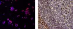
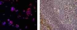
- Immunocytochemistry of fixed and permeabilized HepG2 cells using 1 µg/mL of Anti-Human/Mouse Sox9 Purified followed by 1 µg/mL of F (ab')2 Anti-Mouse IgG eFluor® 570.Nuclei are counterstained with DAPI, and colocalization of signal appears pink (left).Immunohistochemistry of formalin-fixed paraffin embedded human tonsil using 1 µg/mL of Anti-Human/Mouse Sox9 Purified followed by Anti-Mouse IgG Biotin, Streptavidin HRP, and DAB visualization.Nuclei are counterstained with hematoxylin (right).

- Submitted by
- Invitrogen Antibodies (provider)
- Main image
- Experimental details
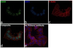
- Immunofluorescence analysis of SOX9 was performed using 70% confluent log phase HCT 116 cells. The cells were fixed with 4% paraformaldehyde for 10 minutes, permeabilized with 0.1% Triton™ X-100 for 15 minutes, and blocked with 2% BSA for 45 minutes at room temperature. The cells were labeled with SOX9 Monoclonal Antibody (GMPR9), eBioscience™ (Product # 14-9765-82) at 1 µg/mL in 0.1% BSA, incubated at 4 degree celsius overnight and then labeled with Donkey anti-Mouse IgG (H+L) Highly Cross-Adsorbed Secondary Antibody, Alexa Fluor Plus 488 (Product # A32766, 1:2000 dilution), for 45 minutes at room temperature (Panel a: Green). Nuclei (Panel b: Blue) were stained with ProLong™ Diamond Antifade Mountant with DAPI (Product # P36962). F-actin (Panel c: Red) was stained with Rhodamine Phalloidin (Product # R415, 1:300 dilution). Panel d represents the merged image showing nuclear localization. Panel e represents control cells with no primary antibody to assess background. The images were captured at 60X magnification.

- Submitted by
- Invitrogen Antibodies (provider)
- Main image
- Experimental details
- Immunocytochemistry of fixed and permeabilized HepG2 cells using 1 µg/mL of Anti-Human/Mouse Sox9 Purified followed by 1 µg/mL of F (ab')2 Anti-Mouse IgG eFluor® 570.Nuclei are counterstained with DAPI, and colocalization of signal appears pink (left).Immunohistochemistry of formalin-fixed paraffin embedded human tonsil using 1 µg/mL of Anti-Human/Mouse Sox9 Purified followed by Anti-Mouse IgG Biotin, Streptavidin HRP, and DAB visualization.Nuclei are counterstained with hematoxylin (right).

- Submitted by
- Invitrogen Antibodies (provider)
- Main image

- Experimental details
- Immunofluorescence analysis of SOX9 was performed using 70% confluent log phase HCT 116 cells. The cells were fixed with 4% paraformaldehyde for 10 minutes, permeabilized with 0.1% Triton™ X-100 for 15 minutes, and blocked with 2% BSA for 45 minutes at room temperature. The cells were labeled with SOX9 Monoclonal Antibody (GMPR9), eBioscience™ (Product # 14-9765-82) at 1 µg/mL in 0.1% BSA, incubated at 4 degree celsius overnight and then labeled with Donkey anti-Mouse IgG (H+L) Highly Cross-Adsorbed Secondary Antibody, Alexa Fluor Plus 488 (Product # A32766, 1:2000 dilution), for 45 minutes at room temperature (Panel a: Green). Nuclei (Panel b: Blue) were stained with ProLong™ Diamond Antifade Mountant with DAPI (Product # P36962). F-actin (Panel c: Red) was stained with Rhodamine Phalloidin (Product # R415, 1:300 dilution). Panel d represents the merged image showing nuclear localization. Panel e represents control cells with no primary antibody to assess background. The images were captured at 60X magnification.

Supportive validation
- Submitted by
- Invitrogen Antibodies (provider)
- Main image
- Experimental details
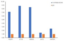
- Chromatin Immunoprecipitation (ChIP) assay of endogenous SOX9 protein using SOX9 Antibody: ChIP was performed using Anti-SOX9 Monoclonal Antibody (GMPR9), eBioscience (Product # 14-9765-82, 2.5 µg) on sheared chromatin from HCT 116 cells using the MAGnify ChIP System kit (Product # 49-2024). Normal Mouse IgG was used as a negative IP control. The purified DNA was analyzed by qPCR using primers binding to RLBP1, ETV2 and RBE65 (Active) and GAPDH and SAT2 satellite repeats (Inactive). Data is presented as fold enrichment of the antibody signal versus the negative control IgG using the comparative CT method.

- Submitted by
- Invitrogen Antibodies (provider)
- Main image

- Experimental details
- Chromatin Immunoprecipitation (ChIP) assay of endogenous SOX9 protein using SOX9 Antibody: ChIP was performed using Anti-SOX9 Monoclonal Antibody (GMPR9), eBioscience (Product # 14-9765-82, 2.5 µg) on sheared chromatin from HCT 116 cells using the MAGnify ChIP System kit (Product # 49-2024). Normal Mouse IgG was used as a negative IP control. The purified DNA was analyzed by qPCR using primers binding to RLBP1, ETV2 and RBE65 (Active) and GAPDH and SAT2 satellite repeats (Inactive). Data is presented as fold enrichment of the antibody signal versus the negative control IgG using the comparative CT method.
