 Explore
 Validate
 Learn
Learn Western blot
Antibody data
- Antibody Data
- Antigen structure
- References [3]
- Comments [0]
- Validations
- Western blot [3]
- Immunocytochemistry [1]
- Immunohistochemistry [6]
- Other assay [3]
Submit
Validation data
Reference
Comment
Report error
- Product number
- PA5-81966 - Provider product page

- Provider
- Invitrogen Antibodies
- Product name
- SOX9 Polyclonal Antibody
- Antibody type
- Polyclonal
- Antigen
- Recombinant full-length protein
- Description
- Immunogen sequence: SQRTHIKTEQ LSPSHYSEQQ QHSPQQIAYS PFNLPHYSPS YPPITRSQYD YTDHQNSSSY YSHAAGQGTG LYSTFTYMNP AQRPMYTPIA DTSGVPSIPQ THSPQHWEQP VYTQLTR
- Reactivity
- Human, Mouse, Rat
- Host
- Rabbit
- Isotype
- IgG
- Vial size
- 100 µL
- Concentration
- 0.08 mg/mL
- Storage
- Store at 4°C short term. For long term storage, store at -20°C, avoiding freeze/thaw cycles.
Submitted references Connexin32 regulates expansion of liver cancer stem cells via the PI3K/Akt signaling pathway.
Derivation and Characterization of Murine and Amphibian Müller Glia Cell Lines.
Survival and cellular heterogeneity of epithelium in cultured mouse and rat precision-cut intestinal slices.
Li H, Wang B, Qi B, Jiang G, Qin M, Yu M
Oncology reports 2022 Sep;48(3)
Oncology reports 2022 Sep;48(3)
Derivation and Characterization of Murine and Amphibian Müller Glia Cell Lines.
Gallo RA, Qureshi F, Strong TA, Lang SH, Pino KA, Dvoriantchikova G, Pelaez D
Translational vision science & technology 2022 Apr 1;11(4):4
Translational vision science & technology 2022 Apr 1;11(4):4
Survival and cellular heterogeneity of epithelium in cultured mouse and rat precision-cut intestinal slices.
Biel C, Bigaeva E, Hesse M, Bomers JJM, van Summeren K, Teunis MAT, Vaessen S, Ten Klooster JP, Olinga P
Toxicology in vitro : an international journal published in association with BIBRA 2020 Dec;69:104974
Toxicology in vitro : an international journal published in association with BIBRA 2020 Dec;69:104974
No comments: Submit comment
Supportive validation
- Submitted by
- Invitrogen Antibodies (provider)
- Main image

- Experimental details
- Western blot analysis of SOX9 by a SOX9 polyclonal antibody (Product # PA5-81966). Analysis in human cell line HepG2.

- Submitted by
- Invitrogen Antibodies (provider)
- Main image

- Experimental details
- Western blot analysis of SOX9 by a SOX9 polyclonal antibody (Product # PA5-81966). Analysis in mouse cell line NIH-3T3 and rat cell line NBT-II.

- Submitted by
- Invitrogen Antibodies (provider)
- Main image
- Experimental details
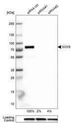
- Western blot analysis of SOX9 by a SOX9 polyclonal antibody (Product # PA5-81966). Analysis in U-251MG cells transfected with control siRNA, target specific siRNA probe #1 and #2, using Anti-SOX9.

Supportive validation
- Submitted by
- Invitrogen Antibodies (provider)
- Main image

- Experimental details
- Immunofluorescent analysis of SOX9 in U-251 MG cells using a SOX9 polyclonal antibody (Product # PA5-81966). The analysis shows localization to nucleoplasm.

Supportive validation
- Submitted by
- Invitrogen Antibodies (provider)
- Main image

- Experimental details
- Immunohistochemical analysis of SOX9 in human testis using a SOX9 polyclonal antibody (Product # PA5-81966). The analysis shows moderate nuclear positivity in a subset of cells in seminiferous ducts.

- Submitted by
- Invitrogen Antibodies (provider)
- Main image

- Experimental details
- Immunohistochemical analysis of SOX9 in human glioma using a SOX9 polyclonal antibody (Product # PA5-81966). The analysis shows moderate to strong nuclear positivity in tumor cells.

- Submitted by
- Invitrogen Antibodies (provider)
- Main image

- Experimental details
- Immunohistochemical analysis of SOX9 in human colorectal cancer using a SOX9 polyclonal antibody (Product # PA5-81966). The analysis shows moderate to strong nuclear positivity in tumor cells.

- Submitted by
- Invitrogen Antibodies (provider)
- Main image

- Experimental details
- Immunohistochemical analysis of SOX9 in human small intestine using a SOX9 polyclonal antibody (Product # PA5-81966). The analysis shows moderate nuclear positivity in a subset of glandular cells.

- Submitted by
- Invitrogen Antibodies (provider)
- Main image

- Experimental details
- Immunohistochemical analysis of SOX9 in human skeletal muscle using a SOX9 polyclonal antibody (Product # PA5-81966). The analysis shows no nuclear positivity in striated muscle fibers as expected.

- Submitted by
- Invitrogen Antibodies (provider)
- Main image

- Experimental details
- Immunohistochemical analysis of SOX9 in human testis and skeletal muscle tissues using a SOX9 polyclonal antibody (Product # PA5-81966). Corresponding RNA-seq data are presented for the same tissues.

Supportive validation
- Submitted by
- Invitrogen Antibodies (provider)
- Main image

- Experimental details
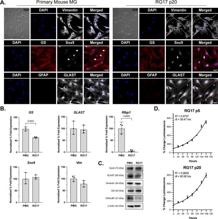
- Figure 1. Immunophenotypic characterization of mouse MG cell line RG17. (A) Phase contrast images and immunofluorescence staining of primary MG cells and passage 20 RG17 cells. RG17 demonstrate typical MG morphology in culture and express many well-characterized MG markers. Scale bar, 50 um and applies to all immunofluorescence images. (B) Expression of characteristic MG genes in primary MG and RG17 cells (passages 20-25). The geometric means and geometric standard deviations ( N = 3 independent cultures) are graphed. An unpaired t -test was applied with a P value of
- Submitted by
- Invitrogen Antibodies (provider)
- Main image

- Experimental details
- Figure 2. Immunophenotypic characterization of X laevis MG cell line XG69. (A) Phase contrast images and immunofluorescence staining of primary MG and XG69 cell line at passage 20. (B) Expression of MG genes in primary MG and XG69 cells (passages 20-25). The geometric means and geometric standard deviations ( N = 3 independent cultures) are graphed. An unpaired t -test was applied with a P value of

- Submitted by
- Invitrogen Antibodies (provider)
- Main image

- Experimental details
- Figure 4. Cx32 regulates the expansion of LCSCs. (A) Effect of Cx32 knockdown on the mRNA expression levels of stemness-associated genes in HepG2 cells. (B) Effect of Cx32 overexpression on the mRNA expression levels of stemness-associated genes in HCCLM3 cells. (C and D) The expression levels of stemness-associated genes were examined by western blotting when HepG2 cells were transfected with shRNA-Cx32. The proteins were normalized with beta-actin. (E and F) The expression levels of stemness-associated genes were observed when HCCLM3 cells overexpressed Cx32. The proteins were normalized with beta-actin. (G and H) Effect of Cx32 knockdown on the sphere-forming capacities of HepG2 cells. Magnification, x200. The bar graph shows the average number of spheres >100 um in diameter. (I and J) Effect of Cx32 overexpression on the sphere-forming capacities of HCCLM3 cells. Magnification, x200. The bar graph shows the average number of spheres >100 um in diameter. For the aforementioned images, the error bars represent the mean +- SEM of three independent experiments; *P
