 Explore
 Validate
 Learn
LearnMA1-16512
antibody from Invitrogen Antibodies
Targeting: ARNT
bHLHe2, HIF-1beta
 Western blot Immunocytochemistry
 Immunoprecipitation Immunohistochemistry Gel shift Chromatin Immunoprecipitation
Antibody data
- Antibody Data
- Antigen structure
- References [2]
- Comments [0]
- Validations
- Western blot [3]
- Immunocytochemistry [1]
- Immunohistochemistry [1]
Submit
Validation data
Reference
Comment
Report error
- Product number
- MA1-16512 - Provider product page

- Provider
- Invitrogen Antibodies
- Product name
- HIF-1 beta Monoclonal Antibody (H1beta234)
- Antibody type
- Monoclonal
- Antigen
- Other
- Description
- Suggested positive control: antigen standard for ARNT (transient overexpression lysate).
- Reactivity
- Human, Mouse, Rat, Bovine
- Host
- Mouse
- Isotype
- IgG
- Antibody clone number
- H1beta234
- Vial size
- 100 µL
- Concentration
- 1 mg/mL
- Storage
- -20° C, Avoid Freeze/Thaw Cycles
Submitted references HIF-1alpha-targeted pathways are activated by heat acclimation and contribute to acclimation-ischemic cross-tolerance in the heart.
Identification of functional hypoxia response elements in the promoter region of the DEC1 and DEC2 genes.
Maloyan A, Eli-Berchoer L, Semenza GL, Gerstenblith G, Stern MD, Horowitz M
Physiological genomics 2005 Sep 21;23(1):79-88
Physiological genomics 2005 Sep 21;23(1):79-88
Identification of functional hypoxia response elements in the promoter region of the DEC1 and DEC2 genes.
Miyazaki K, Kawamoto T, Tanimoto K, Nishiyama M, Honda H, Kato Y
The Journal of biological chemistry 2002 Dec 6;277(49):47014-21
The Journal of biological chemistry 2002 Dec 6;277(49):47014-21
No comments: Submit comment
Supportive validation
- Submitted by
- Invitrogen Antibodies (provider)
- Main image

- Experimental details
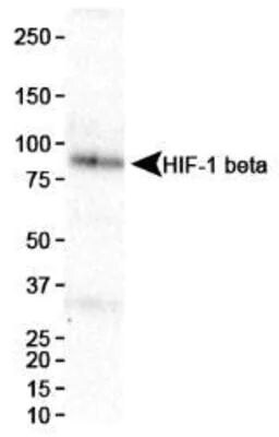
- Western blot analysis of HIF-1 beta in HeLa nuclear extract. Samples were incubated in HIF-1 beta monoclonal antibody (Product # MA1-16512). Theoretical molecular weight 86.6 kDa. Observed molecular weight ~85 kDa.
- Submitted by
- Invitrogen Antibodies (provider)
- Main image

- Experimental details
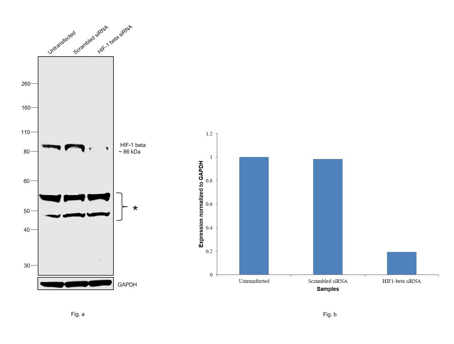
- Knockdown of HIF-1 beta was achieved by transfecting HeLa with HIF-1 beta specific siRNAs (Silencer® select Product # s1613, s1614). Western blot analysis (Fig. a) was performed using whole cell extracts from the HIF-1 beta knockdown cells (lane 3), non-specific scrambled siRNA transfected cells (lane 2) and untransfected cells (lane 1). The blot was probed with HIF-1 beta Monoclonal Antibody (H1beta234) (Product # MA1-16512, 1 µg/mL) and Goat anti-Mouse IgG (H+L) Superclonal™ Recombinant Secondary Antibody, HRP (Product # A28177, 1:4000 dilution). Densitometric analysis of this western blot is shown in histogram (Fig. b). Decrease in signal upon siRNA mediated knock down confirms that antibody is specific to HIF-1 beta.
- Submitted by
- Invitrogen Antibodies (provider)
- Main image
- Experimental details
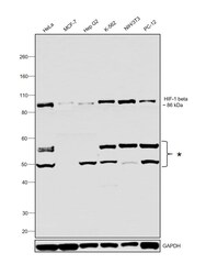
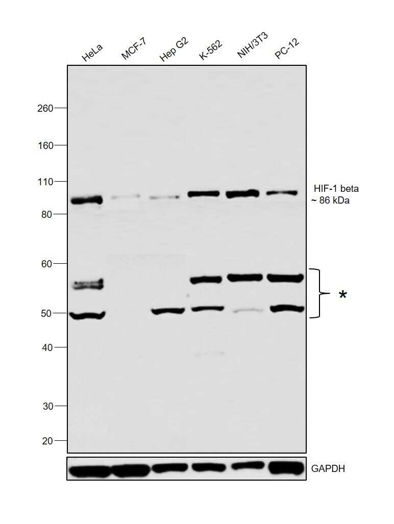
- Western blot was performed using Anti-HIF-1 beta Monoclonal Antibody (H1beta234) (Product # MA1-16512) and a 86 kDa band corresponding to HIF-1 beta was observed across cell lines tested. Additional uncharacterized bands (*) were observed between 50-60 kDa. Whole cell extracts (30 µg lysate) of HeLa (Lane 1), MCF-7 (Lane 2), Hep G2 (Lane 3), K-562 (Lane 4), NIH/3T3 (Lane 5) and PC-12 (Lane 6) were electrophoresed using NuPAGE™ 4-12% Bis-Tris Protein Gel (Product # NP0322BOX). Resolved proteins were then transferred onto a nitrocellulose membrane (Product # IB23001) by iBlot® 2 Dry Blotting System (Product # IB21001). The blot was probed with the primary antibody (1 µg/mL) and detected by chemiluminescence with Goat anti-Mouse IgG (H+L) Superclonal™ Recombinant Secondary Antibody, HRP (Product # A28177, 1:4000 dilution) using the iBright FL 1000 (Product # A32752). Chemiluminescent detection was performed using Novex® ECL Chemiluminescent Substrate Reagent Kit (Product # WP20005).
Supportive validation
- Submitted by
- Invitrogen Antibodies (provider)
- Main image
- Experimental details
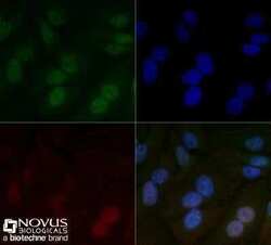
- Immunocytochemistry analysis of HIF-1 beta in HeLa cells fixed for 10 minutes using 10% formalin and then permeabilized for 5 minutes using 1X TBS + 0.5% Triton X-100. Samples were incubated in HIF-1 beta monoclonal antibody (Product # MA1-16512) using a dilution of 5 µg/mL overnight at 4 °C followed by anti-mouse DyLight 488 (Green) with a dilution of 1:500. Actin was detected with Phalloidin 568 (Red) at a 1:200 dilution. Nuclei were counterstained with DAPI (Blue). Cells were imaged using a 40X objective.

Supportive validation
- Submitted by
- Invitrogen Antibodies (provider)
- Main image
- Experimental details
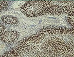
- Immunohistochemical analysis of HIF-1 beta in human glioblastoma multi-forme. Samples were incubated in HIF-1 beta monoclonal antibody (Product # MA1-16512).
