Explore
 Validate
 Learn
LearnAP32519PU-N
antibody from Acris Antibodies GmbH
Targeting: OLIG2
BHLHB1, bHLHe19, OLIGO2, PRKCBP2, RACK17
 Western blot
Antibody data
- Antibody Data
- Antigen structure
- References [0]
- Comments [0]
- Validations
- Western blot [1]
- Immunocytochemistry [1]
- Immunoprecipitation [1]
- Immunohistochemistry [2]
Submit
Validation data
Reference
Comment
Report error
- Product number
- AP32519PU-N - Provider product page

- Provider
- Acris Antibodies GmbH
- Product name
- anti OLIG2
- Antibody type
- Polyclonal
- Antigen
- Synthetic peptide
- Reactivity
- Human, Mouse, Rat
- Host
- Rabbit
- Vial size
- 0.1 mg
- Concentration
- 1.0 mg/ml
No comments: Submit comment
Supportive validation
- Submitted by
- Acris Antibodies GmbH (provider)
- Main image
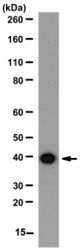
- Experimental details
- Western blot analysis with mouse brain tissue lysate using OLIG2 antibody Cat.-No. AP32519PU-N (Lot 2172341, 1 μg/ml dilution), HRP conjugated donkey anti-rabbit IgG secondary antibody and a chemiluminescence detection system. Arrow indicates OLIG2 (~37 kDa). (Representative lot data).

Supportive validation
- Submitted by
- Acris Antibodies GmbH (provider)
- Main image
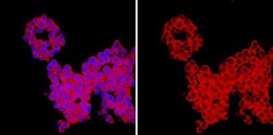
- Experimental details
- Immunocytochemistry: confocal fluorescent analysis of PC12 cells using OLIG2 antibody Cat.-No. AP32519PU-N (Lot 2179341) (Red). Nucleus is stained with DAPI (Blue). This antibody positively stains the cytoplasm. (Representative lot data.)

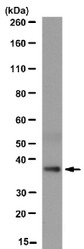
Supportive validation
- Submitted by
- Acris Antibodies GmbH (provider)
- Main image

- Experimental details
- Immunoprecipitation analysis: 10 μg of OLIG2 antibody Cat.-No. AP32519PU-N (Lot 2179341) immunoprecipitated OLIG2 (~37 kDa) from 500μg of mouse brain RIPA lysate. (Representative lot data.)
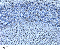
Supportive validation
- Submitted by
- Acris Antibodies GmbH (provider)
- Main image
- Experimental details
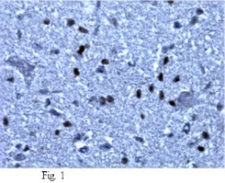
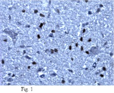
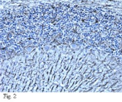
- Immunohistochemical analysis on Paraffin-embedded normal rat hindbrain (Fig. 1) and normal rat cerebellum (Fig. 2, see below) tissues (heat-induced epitope retrieval in citrate buffer, pH 6.0) using a 1:500 dilution of OLIG2 antibody Cat. No. AP32519PU-N (Lot 2179341). Reactivity was detected using the IHCSelect® Detection Kit. In Fig. 2, GFP staining was used as a positive control to determine the sucess of tissue fixation and antigen retrieval. Staining pattern in Fig. 1, appears to be restricted to the cytoplasm and nucleus. (Representative lot data).
- Submitted by
- Acris Antibodies GmbH (provider)
- Main image
- Experimental details
- IHC on Paraffin-embedded normal rat cerebellum using OLIG2 antibody Cat.-No. AP32519PU-N (further explanations see IHC image Fig. 1').