 Explore
 Validate
 Learn
Learn Western blot
Antibody data
- Antibody Data
- Antigen structure
- References [1]
- Comments [0]
- Validations
- Western blot [1]
- Immunocytochemistry [2]
- Immunohistochemistry [5]
Submit
Validation data
Reference
Comment
Report error
- Product number
- HPA003258 - Provider product page

- Provider
- Atlas Antibodies
- Proper citation
- Atlas Antibodies Cat#HPA003258, RRID:AB_1078928
- Product name
- Anti-GABPA
- Antibody type
- Polyclonal
- Description
- Polyclonal Antibody against Human GABPA, Gene description: GA binding protein transcription factor, alpha subunit 60kDa, Alternative Gene Names: E4TF1-60, E4TF1A, NFT2, NRF2, NRF2A, Validated applications: ICC, IHC, WB, Uniprot ID: Q06546, Storage: Store at +4°C for short term storage. Long time storage is recommended at -20°C.
- Reactivity
- Human
- Host
- Rabbit
- Conjugate
- Unconjugated
- Isotype
- IgG
- Vial size
- 100 µl
- Concentration
- 0.1 mg/ml
- Storage
- Store at +4°C for short term storage. Long time storage is recommended at -20°C.
Submitted references The ETS family member GABPα modulates androgen receptor signalling and mediates an aggressive phenotype in prostate cancer
Sharma N, Massie C, Butter F, Mann M, Bon H, Ramos-Montoya A, Menon S, Stark R, Lamb A, Scott H, Warren A, Neal D, Mills I
Nucleic Acids Research 2014;42(10):6256-6269
Nucleic Acids Research 2014;42(10):6256-6269
No comments: Submit comment
Supportive validation
- Submitted by
- Atlas Antibodies (provider)
- Main image

- Experimental details
- Western blot analysis in human cell line Daudi.
- Sample type
- Human
- Protocol
- Protocol

Supportive validation
- Submitted by
- Atlas Antibodies (provider)
- Main image

- Experimental details
- Immunofluorescent staining of human cell line U-251 MG shows localization to nucleoplasm.
- Sample type
- HUMAN

- Submitted by
- Atlas Antibodies (provider)
- Main image

- Experimental details
- Immunofluorescent staining of human cell line U-251 MG shows localization to nucleoplasm.
- Sample type
- Human
- Protocol
- Protocol

Supportive validation
- Submitted by
- Atlas Antibodies (provider)
- Main image
- Experimental details
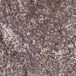
- Immunohistochemical staining of human lymph node shows strong nuclear positivity in non-germinal center cells.
- Sample type
- HUMAN

- Submitted by
- Atlas Antibodies (provider)
- Main image

- Experimental details
- Immunohistochemical staining of human placenta shows strong nuclear positivity in trophoblastic cells.
- Sample type
- Human
- Protocol
- Protocol

- Submitted by
- Atlas Antibodies (provider)
- Main image

- Experimental details
- Immunohistochemical staining of human tonsil shows strong nuclear positivity in non-germinal center cells.
- Sample type
- Human
- Protocol
- Protocol

- Submitted by
- Atlas Antibodies (provider)
- Main image

- Experimental details
- Immunohistochemical staining of human skin shows strong nuclear positivity in squamous epithelial cells.
- Sample type
- Human
- Protocol
- Protocol

- Submitted by
- Atlas Antibodies (provider)
- Main image

- Experimental details
- Immunohistochemical staining of human cerebellum shows no positivity in Purkinje cells as expected.
- Sample type
- Human
- Protocol
- Protocol
