Explore
 Validate
 Learn
Learn Western blot
 Immunoprecipitation
Antibody data
- Antibody Data
- Antigen structure
- References [2]
- Comments [0]
- Validations
- Western blot [4]
- Immunohistochemistry [3]
- Other assay [2]
Submit
Validation data
Reference
Comment
Report error
- Product number
- PA5-19956 - Provider product page

- Provider
- Invitrogen Antibodies
- Product name
- RIP3 Polyclonal Antibody
- Antibody type
- Polyclonal
- Antigen
- Synthetic peptide
- Reactivity
- Human, Mouse, Rat
- Host
- Rabbit
- Isotype
- IgG
- Vial size
- 100 µg
- Concentration
- 1 mg/mL
- Storage
- 4° C
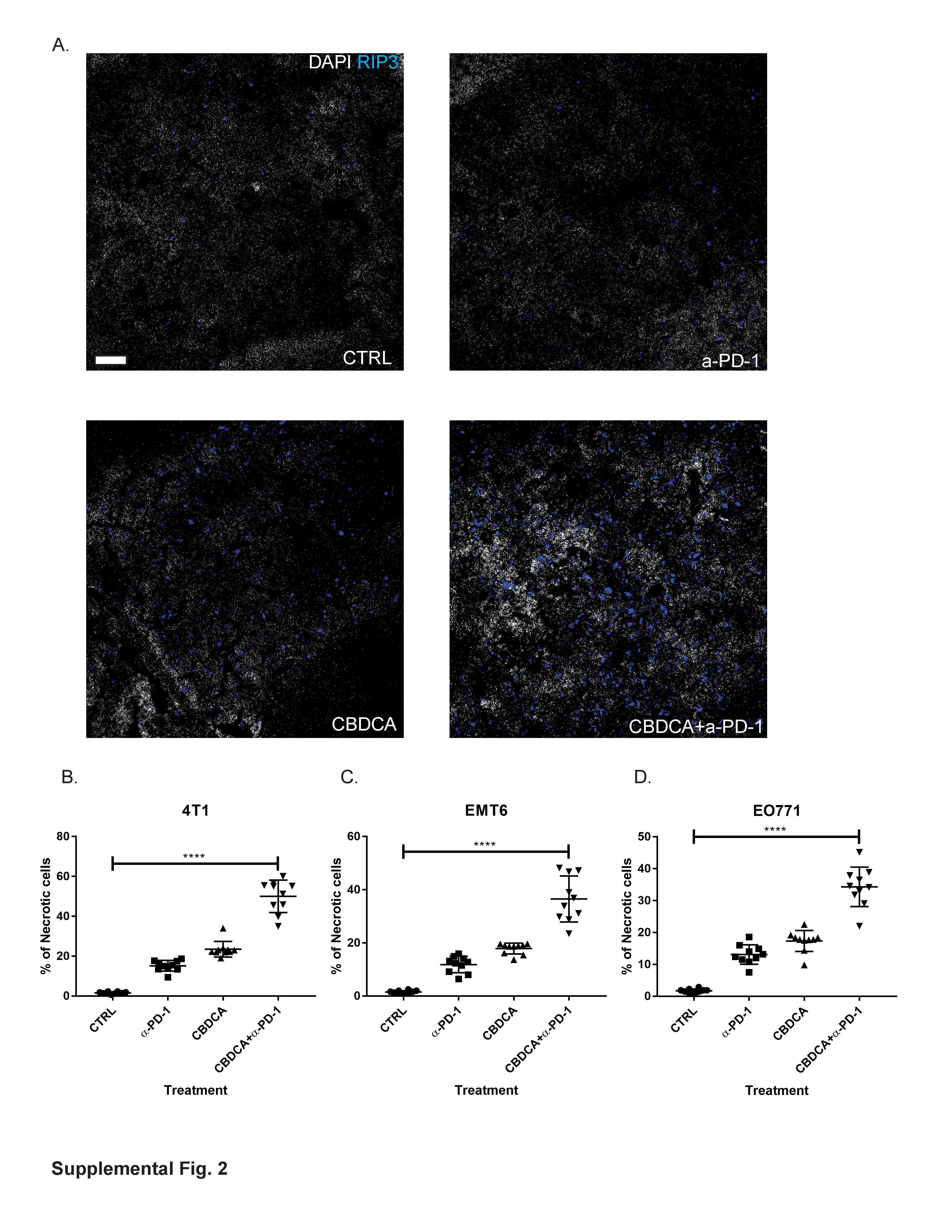
Submitted references Therapy With Carboplatin and Anti-PD-1 Antibodies Before Surgery Demonstrates Sustainable Anti-Tumor Effects for Secondary Cancers in Mice With Triple-Negative Breast Cancer.
Autophosphorylation at serine 166 regulates RIP kinase 1-mediated cell death and inflammation.
Gao M, Wang T, Ji L, Bai S, Tian L, Song H
Frontiers in immunology 2020;11:366
Frontiers in immunology 2020;11:366
Autophosphorylation at serine 166 regulates RIP kinase 1-mediated cell death and inflammation.
Laurien L, Nagata M, Schünke H, Delanghe T, Wiederstein JL, Kumari S, Schwarzer R, Corona T, Krüger M, Bertrand MJM, Kondylis V, Pasparakis M
Nature communications 2020 Apr 8;11(1):1747
Nature communications 2020 Apr 8;11(1):1747
No comments: Submit comment
Supportive validation
- Submitted by
- Invitrogen Antibodies (provider)
- Main image
- Experimental details
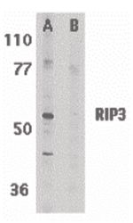
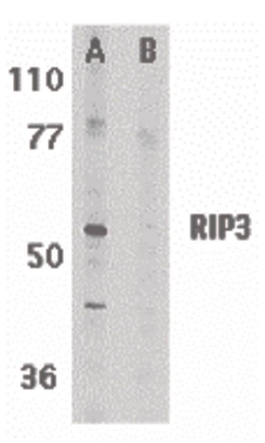
- Western blot analysis of mouse 3T3 whole cell lysate in the absence (A) or presence (B) of blocking peptide using a RIP3 polyclonal antibody (Product # PA5-19956) at 1 µg/mL.
- Submitted by
- Invitrogen Antibodies (provider)
- Main image
- Experimental details
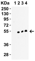
- Western Blot Validation in C2C12 Cells Mouse. Loading: 15 µg of lysates per lane. Antibodies: RIP3 Polyclonal Antibody (Product # PA5-19956) 1h incubation at RT in 0.05 NFDM/TBST. Secondary: Goat anti-rabbit IgG HRP conjugate at 1:10,000 dilution. Lane 1: RIP3 Polyclonal Antibody, 0.1 µg/mL in the presence of peptide blocking Lane 2: RIP3 Polyclonal Antibody, 0.1 µg/mL Lane 3: RIP3 Polyclonal Antibody, 0.2 µg/mL Lane 4: RIP3 Polyclonal Antibody, 0.5 µg/mL

- Submitted by
- Invitrogen Antibodies (provider)
- Main image
- Experimental details
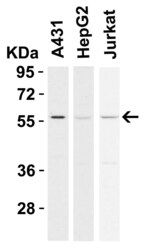
- Western Blot Validation in Human Cell Lines. Loading: 15 µg of lysates per lane. Antibodies: RIP3 Polyclonal Antibody (Product # PA5-19956) (0.5 µg/mL), 1h incubation at RT in 0.05 NFDM/TBST. Secondary: Goat anti-rabbit IgG HRP conjugate at 1:10,000 dilution.

- Submitted by
- Invitrogen Antibodies (provider)
- Main image
- Experimental details
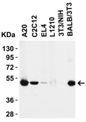
- Western Blot Validation in Mouse Cell lines. Loading: 15 µg of lysates per lane. Antibodies: RIP3 Polyclonal Antibody (Product # PA5-19956) (0.5 µg/mL), 1h incubation at RT in 0.05 NFDM/TBST. Secondary: Goat anti-rabbit IgG HRP conjugate at 1:10,000 dilution.

Supportive validation
- Submitted by
- Invitrogen Antibodies (provider)
- Main image
- Experimental details
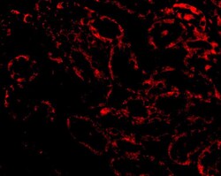
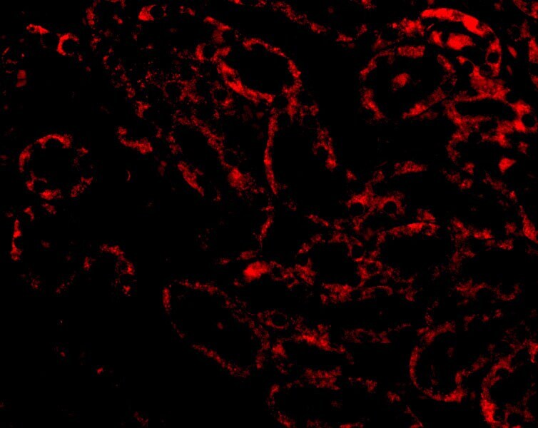
- Immunofluorescent analysis of 4% paraformaldehyde-fixed Rat Kidney tissue labeling RIP3 with RIP3 Polyclonal Antibody (Product # PA5-19956) at 20 µg/mL, followed by goat anti-rabbit IgG secondary antibody at 1:500 dilution (red).
- Submitted by
- Invitrogen Antibodies (provider)
- Main image
- Experimental details
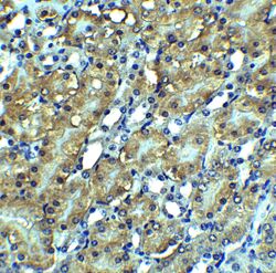
- Immunohistochemical analysis of paraffin-embedded mouse kidney tissue using RIP3 Polyclonal Antibody (Product # PA5-19956) at 2.5 µg/mL. Tissue was fixed with formaldehyde and blocked with 0.1 serum for 1 h at RT; antigen retrieval was by heat mediation with a citrate buffer (pH6). Samples were incubated with primary antibody overnight at 4˚C. A goat anti-rabbit IgG H&L (HRP) at 1/250 was used as secondary. Counter stained with Hematoxylin.

- Submitted by
- Invitrogen Antibodies (provider)
- Main image
- Experimental details
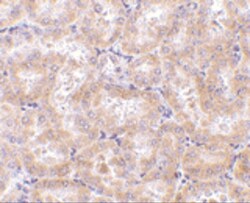
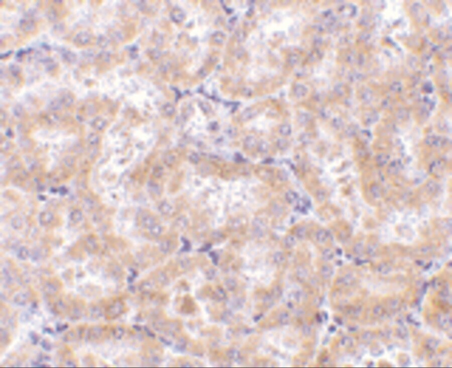
- Immunohistochemical analysis of paraffin-embedded rat kidney tissue using RIP3 Polyclonal Antibody (Product # PA5-19956) at 5 µg/mL. Tissue was fixed with formaldehyde and blocked with 0.1 serum for 1 h at RT; antigen retrieval was by heat mediation with a citrate buffer (pH6). Samples were incubated with primary antibody overnight at 4˚C. A goat anti-rabbit IgG H&L (HRP) at 1/250 was used as secondary. Counter stained with Hematoxylin.
Supportive validation
- Submitted by
- Invitrogen Antibodies (provider)
- Main image

- Experimental details
- NULL
- Submitted by
- Invitrogen Antibodies (provider)
- Main image
- Experimental details
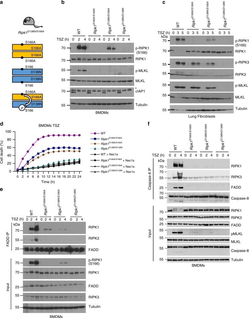
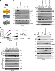
- Fig. 7 S166 phosphorylation enhances RIPK1 kinase activity. a Scheme depicting the possible combinations of RIPK1 dimers in Ripk1 D138N/S166A mice. Residue 166 on RIPK1 is indicated. b , BMDMs from mice of the indicated genotypes were treated with a combination of TNF (T; 10 ng mL -1 ), SMAC mimetic (S (Birinapant), 1 muM) and Z-VAD-FMK (Z) for 0, 2 or 4 h and lysates were analyzed by immunoblot with the indicated antibodies. Blots shown are representative of three independent experiments. *Non-specific band. c Primary lung fibroblasts from mice of the indicated genotypes were treated with a combination of TNF (T; 20 ng mL -1 ), SMAC mimetic (S (Birinapant), 1 muM) and Z-VAD-FMK (Z) for 0, 3 or 5 h and lysates were analyzed by immunoblot with the indicated antibodies. Blots shown are representative of three independent experiments. d BMDMs from mice of the indicated genotypes were treated with combinations of TNF (T; 10 ng mL -1 ), SMAC mimetic (S (Briniapant), 1 muM) and Z-VAD-FMK (Z) in the presence or absence of Nec-1s. Cell death was measured using IncuCyte. Graph shows mean of technical triplicates. Data are representative of three independent experments. For purpose of comparability the data of the genotypes WT, Ripk1 S166A/S166A and Ripk1 D138N/D138N shown here are identical to those in Fig. 1b . e BMDMs were treated with a combination of TNF (T; 10 ng mL -1 ), SMAC mimetic (S (Birinapant), 1 muM) and Z-VAD-FMK (Z) for 0, 2 or 4 h. FADD immunoprecipitates and total cel