Antibody data
- Antibody Data
- Antigen structure
- References [3]
- Comments [0]
- Validations
- Western blot [1]
- Immunocytochemistry [2]
- Immunohistochemistry [1]
- Other assay [1]
Submit
Validation data
Reference
Comment
Report error
- Product number
- PA5-38294 - Provider product page

- Provider
- Invitrogen Antibodies
- Product name
- Anti-MiTF Polyclonal Antibody
- Antibody type
- Polyclonal
- Antigen
- Synthetic peptide
- Reactivity
- Human, Mouse
- Host
- Rabbit
- Isotype
- IgG
- Vial size
- 100 µg
- Concentration
- 1 mg/mL
- Storage
- -20°C
Submitted references D609 protects retinal pigmented epithelium as a potential therapy for age-related macular degeneration.
AKT inhibition-mediated dephosphorylation of TFE3 promotes overactive autophagy independent of MTORC1 in cadmium-exposed bone mesenchymal stem cells.
Stem Cell Derived Retinal Pigment Epithelium: The Role of Pigmentation as Maturation Marker and Gene Expression Profile Comparison with Human Endogenous Retinal Pigment Epithelium.
Wang B, Wang L, Gu S, Yu Y, Huang H, Mo K, Xu H, Zeng F, Xiao Y, Peng L, Liu C, Cao N, Liu Y, Yuan J, Ouyang H
Signal transduction and targeted therapy 2020 Mar 4;5(1):20
Signal transduction and targeted therapy 2020 Mar 4;5(1):20
AKT inhibition-mediated dephosphorylation of TFE3 promotes overactive autophagy independent of MTORC1 in cadmium-exposed bone mesenchymal stem cells.
Pi H, Li M, Zou L, Yang M, Deng P, Fan T, Liu M, Tian L, Tu M, Xie J, Chen M, Li H, Xi Y, Zhang L, He M, Lu Y, Chen C, Zhang T, Wang Z, Yu Z, Gao F, Zhou Z
Autophagy 2019 Apr;15(4):565-582
Autophagy 2019 Apr;15(4):565-582
Stem Cell Derived Retinal Pigment Epithelium: The Role of Pigmentation as Maturation Marker and Gene Expression Profile Comparison with Human Endogenous Retinal Pigment Epithelium.
Bennis A, Jacobs JG, Catsburg LAE, Ten Brink JB, Koster C, Schlingemann RO, van Meurs J, Gorgels TGMF, Moerland PD, Heine VM, Bergen AA
Stem cell reviews and reports 2017 Oct;13(5):659-669
Stem cell reviews and reports 2017 Oct;13(5):659-669
No comments: Submit comment
Supportive validation
- Submitted by
- Invitrogen Antibodies (provider)
- Main image

- Experimental details
- Western blot analysis of MiTF in extracts from HepG2 cells (lane 1) and COLO205 cells (lane 2) using a MiTF polyclonal antibody (Product # PA5-38294).
Supportive validation
- Submitted by
- Invitrogen Antibodies (provider)
- Main image

- Experimental details
- Immunofluorescent analysis of MiTF in HeLa cells using a MiTF polyclonal antibody (Product # PA5-38294).
- Submitted by
- Invitrogen Antibodies (provider)
- Main image

- Experimental details
- Immunofluorescent analysis of MiTF in HeLa cells using a MiTF polyclonal antibody (Product # PA5-38294).
Supportive validation
- Submitted by
- Invitrogen Antibodies (provider)
- Main image

- Experimental details
- Immunohistochemical analysis of MiTF in paraffin-embedded human skin tissue using a MiTF polyclonal antibody (Product # PA5-38294).
Supportive validation
- Submitted by
- Invitrogen Antibodies (provider)
- Main image

- Experimental details
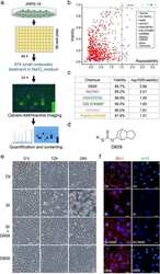
- Fig. 1 High-content screening of oxidative stress inhibitors in ARPE-19 cells. a The workflow of the high-content screening method. b Analysis of the effects on cell viability of each compound by calcein-AM/Hoechst imaging in ARPE-19 cells. R is the value of the distance between the two times of analysis of the viability. c The list of chemical candidates in the library that can inhibit SI-induced cell death in ARPE cells. d Chemical structure of D609. e Phase-contrast images of the ftRPE cells treated with D609 (10 muM), SI (10 muM), or a combination at 0, 12 or 24 h. f Immunofluorescence imaging of ZO-1 and MITF in the ftPRE cells treated with D609, SI, or a combination for 18 h. Scale bar: 100 mum ( e ), 20 mum ( f ). n = 3
 Explore
 Validate
 Learn
Learn Western blot