Explore
 Validate
 Learn
Learn Western blot
Antibody data
- Antibody Data
- Antigen structure
- References [2]
- Comments [0]
- Validations
- Western blot [1]
Submit
Validation data
Reference
Comment
Report error
- Product number
- MAB8935 - Provider product page

- Provider
- R&D Systems
- Product name
- Human Phospho-Smad2/3 (S465/S467) Antibody
- Antibody type
- Monoclonal
- Description
- Protein A or G purified from cell culture supernatant. Detects human Smad2 and Smad3 when dually phosphorylated at S465 and S467 in Western blots.
- Reactivity
- Human
- Host
- Rabbit
- Conjugate
- Unconjugated
- Antigen sequence
NP_005892- Isotype
- IgG
- Antibody clone number
- 1074A
- Vial size
- 100 ug
- Storage
- Use a manual defrost freezer and avoid repeated freeze-thaw cycles. 12 months from date of receipt, -20 to -70 °C as supplied. 1 month, 2 to 8 °C under sterile conditions after reconstitution. 6 months, -20 to -70 °C under sterile conditions after reconstitution.
Submitted references COA-Cl prevented TGF-β1-induced CTGF expression by Akt dephosphorylation in normal human dermal fibroblasts, and it attenuated skin fibrosis in mice models of systemic sclerosis.
Neuronal Protein 3.1 Deficiency Leads to Reduced Cutaneous Scar Collagen Deposition and Tensile Strength due to Impaired Transforming Growth Factor-β1 to -β3 Translation.
Nakai K, Karita S, Igarashi J, Tsukamoto I, Hirano K, Kubota Y
Journal of dermatological science 2019 Apr;94(1):205-212
Journal of dermatological science 2019 Apr;94(1):205-212
Neuronal Protein 3.1 Deficiency Leads to Reduced Cutaneous Scar Collagen Deposition and Tensile Strength due to Impaired Transforming Growth Factor-β1 to -β3 Translation.
Cheng T, Yue M, Aslam MN, Wang X, Shekhawat G, Varani J, Schuger L
The American journal of pathology 2017 Feb;187(2):292-303
The American journal of pathology 2017 Feb;187(2):292-303
No comments: Submit comment
Supportive validation
- Submitted by
- R&D Systems (provider)
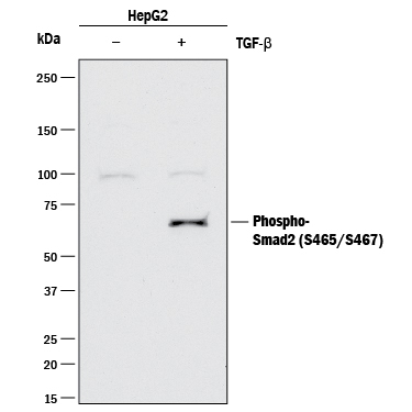
- Main image

- Experimental details
- Detection of Human Phospho-Smad2/3 (S465/S467) by Western Blot. Western blot shows lysates of HepG2 human hepatocellular carcinoma cell line untreated (-) or treated (+) with 10 ng/mL Recombinant Human TGF-beta 1 (Catalog # 240-B) for 30 minutes. PVDF membrane was probed with 2 μg/mL of Rabbit Anti-Human Phospho-Smad2/3 (S465/S467) Monoclonal Antibody (Catalog # MAB8935) followed by HRP-conjugated Anti-Rabbit IgG Secondary Antibody (Catalog # HAF008). A specific band was detected for Phospho-Smad2/3 (S465/S467) at approximately 68 kDa (as indicated). This experiment was conducted under reducing conditions and using Immunoblot Buffer Group 2.