Explore
 Validate
 Learn
Learn Western blot
Antibody data
- Antibody Data
- Antigen structure
- References [0]
- Comments [0]
- Validations
- Western blot [2]
- Immunocytochemistry [1]
Submit
Validation data
Reference
Comment
Report error
- Product number
- PA5-17709 - Provider product page

- Provider
- Invitrogen Antibodies
- Product name
- BCR Polyclonal Antibody
- Antibody type
- Polyclonal
- Antigen
- Synthetic peptide
- Description
- It is not recommended to aliquot this antibody.
- Reactivity
- Human, Mouse, Rat, Porcine
- Host
- Rabbit
- Isotype
- IgG
- Vial size
- 100 µL
- Concentration
- 291 µg/mL
- Storage
- -20°C
No comments: Submit comment
Supportive validation
- Submitted by
- Invitrogen Antibodies (provider)
- Main image

- Experimental details
- KD of BCR was achieved by transfecting Hep G2 with BCR specific siRNAs (Silencer® select Product # s1949). Western blot analysis (Fig. a) was performed using whole cell extracts from the BCR KD cells (Lane 3), non-specific scrambled siRNA transfected cells (Lane 2) and untransfected cells (Lane 1). The blot was probed with BCR Polyclonal Antibody (Product # PA5-17709, 1:1000 dilution) and Goat anti-Rabbit IgG (H+L) Superclonal™ Secondary Antibody, HRP conjugate (Product # A27036, 1:4000 dilution). Densitometric analysis of this western blot is shown in histogram (Fig. b). Loss of signal upon siRNA mediated knock down confirms that antibody is specific to BCR. An uncharacterized band at ~130 kDa was also observed in the samples tested.

- Submitted by
- Invitrogen Antibodies (provider)
- Main image
- Experimental details
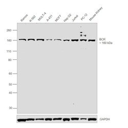
- Western blot was performed using Anti-BCR Polyclonal Antibody (Product # PA5-17709) and a 160 kDa band corresponding to BCR was observed across cell lines and tissue tested. Whole cell extracts (30 µg lysate) of Ramos (Lane 1), K-562 (Lane 2), MOLT-4 (Lane 3), A-431 (Lane 4), MCF7 (Lane 5), Hep G2 (Lane 6), Jurkat (Lane 7), PC-12 (Lane 8) and tissue extract (30 µg lysate) of Mouse Kidney (Lane 9) were electrophoresed using Novex® NuPAGE® 4-12 % Bis-Tris gel (Product # NP0322BOX). Resolved proteins were then transferred onto a nitrocellulose membrane (Product # LC2001) by XCell SureLock™ Mini-Cell and XCell II™ Blot Module (Product # EI0002). The blot was probed with the primary antibody (1:1000 dilution) and detected by chemiluminescence Goat Anti-Rabbit IgG Secondary Antibody, HRP conjugate (Product # A27036, 1:4000 dilution) using the iBright FL 1000 (Product # A32752). Chemiluminescent detection was performed using Novex® ECL Chemiluminescent Substrate Reagent Kit (Product # WP20005).

Supportive validation
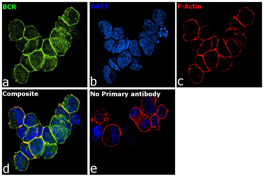
- Submitted by
- Invitrogen Antibodies (provider)
- Main image
- Experimental details
- Immunofluorescence analysis of BCR was performed using K-562 cells. The cells were fixed with 4% paraformaldehyde for 10 minutes, permeabilized with 0.1% Triton™ X-100 for 15 minutes, and blocked with 2% BSA for 1 hour at room temperature. The cells were labeled with BCR Polyclonal Antibody (Product # PA5-17709) at 1:250 dilution in 0.1% BSA, incubated at 4 degree Celsius overnight and then labeled with Goat anti-Rabbit IgG (H+L) Superclonal™ Secondary Antibody, Alexa Fluor® 488 conjugate (Product # A27034) at a dilution of 1:2000 for 45 minutes at room temperature (Panel a: green). Nuclei (Panel b: blue) were stained with ProLong™ Diamond Antifade Mountant with DAPI (Product # P36962). F-actin (Panel c: red) was stained with Rhodamine Phalloidin (Product # R415, 1:300). Panel d represents the merged image showing membranous localization. Panel e represents control cells with no primary antibody to assess background. The images were captured at 60X magnification..