Explore
 Validate
 Learn
Learn Western blot
 Immunoprecipitation
Antibody data
- Antibody Data
- Antigen structure
- References [1]
- Comments [0]
- Validations
- Western blot [3]
- Immunocytochemistry [1]
- Immunohistochemistry [1]
- Other assay [2]
Submit
Validation data
Reference
Comment
Report error
- Product number
- PA5-35984 - Provider product page

- Provider
- Invitrogen Antibodies
- Product name
- SP2 Polyclonal Antibody
- Antibody type
- Polyclonal
- Antigen
- Recombinant protein fragment
- Description
- Recommended positive controls: Jurkat, Raji, NCI-H929, A431, HepG2. Predicted reactivity: Mouse (91%), Rat (90%), Rhesus Monkey (100%), Bovine (93%). Store product as a concentrated solution. Centrifuge briefly prior to opening the vial.
- Reactivity
- Human
- Host
- Rabbit
- Isotype
- IgG
- Vial size
- 100 µL
- Concentration
- 1.65 mg/mL
- Storage
- Store at 4°C short term. For long term storage, store at -20°C, avoiding freeze/thaw cycles.
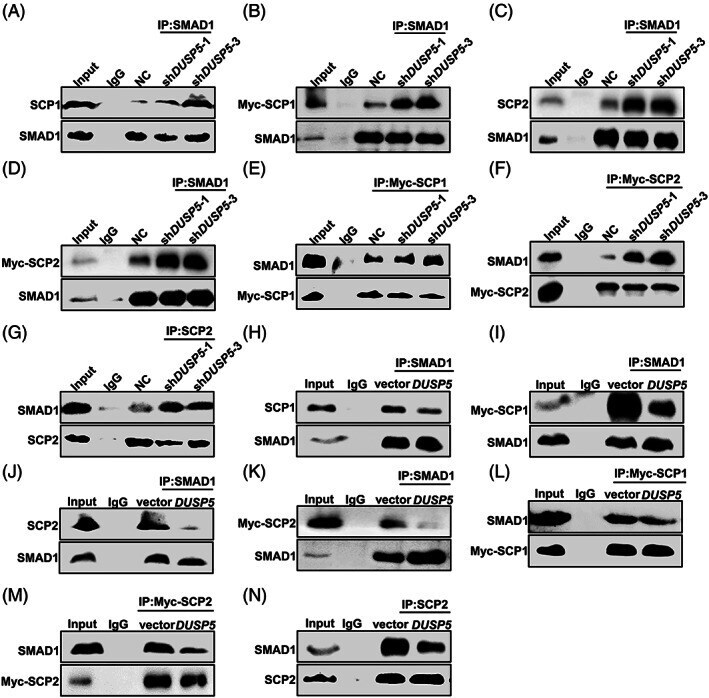
Submitted references DUSP5 promotes osteogenic differentiation through SCP1/2-dependent phosphorylation of SMAD1.
Liu X, Liu X, Du Y, Hu M, Tian Y, Li Z, Lv L, Zhang X, Liu Y, Zhou Y, Zhang P
Stem cells (Dayton, Ohio) 2021 Oct;39(10):1395-1409
Stem cells (Dayton, Ohio) 2021 Oct;39(10):1395-1409
No comments: Submit comment
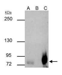
Supportive validation
- Submitted by
- Invitrogen Antibodies (provider)
- Main image
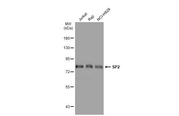
- Experimental details
- Western blot analysis of SP2 using A) 30 µg Jurkat whole cell lysate (B) 30 µg Raji whole cell lysate and C) 30 µg NCI-H929 whole cell lysate. Samples were loaded onto a 7.5% SDS-PAGE gel and probed with a SP2 polyclonal antibody (Product # PA5-35984) at a dilution of 1:500.
- Submitted by
- Invitrogen Antibodies (provider)
- Main image

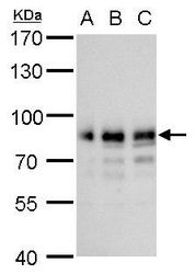
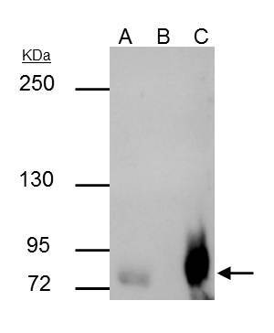
- Experimental details
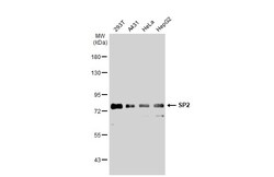
- Western Blot analysis of SP2 was performed by separating 30 µg of various whole cell extracts by 7.5% SDS-PAGE. Proteins were transferred to a membrane and probed with a SP2 Polyclonal Antibody (Product # PA5-35984) at a dilution of 1:1000 and a HRP-conjugated anti-rabbit IgG secondary antibody.
- Submitted by
- Invitrogen Antibodies (provider)
- Main image
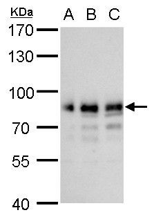
- Experimental details
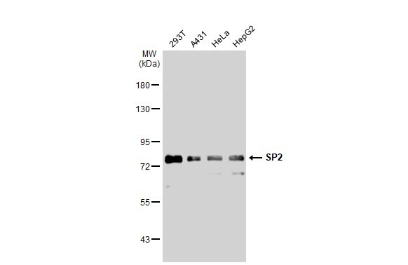
- Western Blot using SP2 Polyclonal Antibody (Product # PA5-35984). Various whole cell extracts (30 µg) were separated by 7.5% SDS-PAGE, and the membrane was blotted with SP2 Polyclonal Antibody (Product # PA5-35984) diluted at 1:1,000. The HRP-conjugated anti-rabbit IgG antibody was used to detect the primary antibody.
Supportive validation
- Submitted by
- Invitrogen Antibodies (provider)
- Main image
- Experimental details
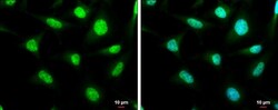
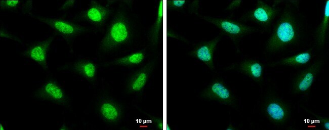
- Immunocytochemistry-Immunofluorescence analysis of SP2 was performed in HeLa cells fixed in 4% paraformaldehyde at RT for 15 min. Green: SP2 Polyclonal Antibody (Product # PA5-35984) diluted at 1:1000. Blue: Hoechst 33342 staining. Scale bar = 10 µm.
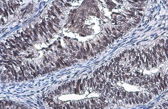
Supportive validation
- Submitted by
- Invitrogen Antibodies (provider)
- Main image

- Experimental details
- Immunohistochemistry (Paraffin) analysis of SP2 was performed in paraffin-embedded human endometrial carcinoma tissue using SP2 Polyclonal Antibody (Product # PA5-35984) at a dilution of 1:500. Antigen Retrieval: Citrate buffer, pH 6.0, 15 min.
Supportive validation
- Submitted by
- Invitrogen Antibodies (provider)
- Main image
- Experimental details
- SP2 Polyclonal Antibody immunoprecipitates SP2 protein in IP experiments. IP Sample: Raji whole cell lysate/extract A : 30 µg whole cell lysate/extract of SP2 protein expressing Raji cells B : Control with 3 µg of pre-immune rabbit IgG C : Immunoprecipitation of SP2 by 3 µg of SP2 Polyclonal Antibody (Product # PA5-35984) 5% SDS-PAGE The immunoprecipitated SP2 protein was detected by SP2 Polyclonal Antibody (Product # PA5-35984) diluted at 1 : 500. Anti-rabbit IgG (HRP) was used as a secondary reagent.
- Submitted by
- Invitrogen Antibodies (provider)
- Main image

- Experimental details
- FIGURE 6 Competitive combination of DUSP5 and SCP1/2 with SMAD1. A-G, Co-IP assay was performed to determine the interaction between SCP1 and SMAD1, and the interaction between SCP2 and SMAD1 in NC, sh DUSP5 -1 and sh DUSP5 -3 HEK293T cells. H-N, The interaction between SCP1 and SMAD1, and the interaction between SCP2 and SMAD1 in vector and DUSP5 HEK293T cells was detected by Co-IP experiment. Data represented were repeated three times. Co-IP, coimmunoprecipitation; DUSP5, dual-specificity phosphatase 5; NC, negative control for sh DUSP5 -1 and sh DUSP5 -3; SCP1, small C-terminal phosphatase 1; SCP2, small C-terminal phosphatase 2; vector, negative control for DUSP5