 Explore
 Validate
 Learn
Learn Western blot
Antibody data
- Antibody Data
- Antigen structure
- References [6]
- Comments [0]
- Validations
- Western blot [3]
- Immunocytochemistry [4]
- Immunohistochemistry [1]
- Flow cytometry [1]
Submit
Validation data
Reference
Comment
Report error
- Product number
- MAB2105 - Provider product page

- Provider
- R&D Systems
- Product name
- Human/Mouse/Rat Vimentin Antibody
- Antibody type
- Monoclonal
- Description
- Protein A or G purified from hybridoma culture supernatant. Vimentin antibodies are ideal for immunocytochemistry colocalization studies in intermediate filaments. Detects human, mouse and rat Vimentin in Western blots.
- Reactivity
- Human, Mouse, Rat
- Host
- Rat
- Conjugate
- Unconjugated
- Antigen sequence
P08670- Isotype
- IgG
- Antibody clone number
- 280618
- Vial size
- 100 ug
- Concentration
- LYOPH
- Storage
- Use a manual defrost freezer and avoid repeated freeze-thaw cycles. 12 months from date of receipt, -20 to -70 °C as supplied. 1 month, 2 to 8 °C under sterile conditions after reconstitution. 6 months, -20 to -70 °C under sterile conditions after reconstitution.
Submitted references Platelet glycoprotein VI and C-type lectin-like receptor 2 deficiency accelerates wound healing by impairing vascular integrity in mice.
RNA sequencing reveals upregulation of a transcriptomic program associated with stemness in metastatic prostate cancer cells selected for taxane resistance.
Acute Drug Effects on the Human Placental Tissue: The Development of a Placental Murine Xenograft Model.
Combined CSL and p53 downregulation promotes cancer-associated fibroblast activation.
GLI1, CTNNB1 and NOTCH1 protein expression in a thymic epithelial malignancy tissue microarray.
Development of a reconstructed cornea from collagen-chondroitin sulfate foams and human cell cultures.
Wichaiyo S, Lax S, Montague SJ, Li Z, Grygielska B, Pike JA, Haining EJ, Brill A, Watson SP, Rayes J
Haematologica 2019 Aug;104(8):1648-1660
Haematologica 2019 Aug;104(8):1648-1660
RNA sequencing reveals upregulation of a transcriptomic program associated with stemness in metastatic prostate cancer cells selected for taxane resistance.
Cajigas-Du Ross CK, Martinez SR, Woods-Burnham L, Durán AM, Roy S, Basu A, Ramirez JA, Ortiz-Hernández GL, Ríos-Colón L, Chirshev E, Sanchez-Hernandez ES, Soto U, Greco C, Boucheix C, Chen X, Unternaehrer J, Wang C, Casiano CA
Oncotarget 2018 Jul 13;9(54):30363-30384
Oncotarget 2018 Jul 13;9(54):30363-30384
Acute Drug Effects on the Human Placental Tissue: The Development of a Placental Murine Xenograft Model.
Verheecke M, Hermans E, Tuyaerts S, Souche E, Van Bree R, Verbist G, Everaert T, Cortès-Calabuig A, Van Houdt J, Van Calsteren K, Amant F
Reproductive sciences (Thousand Oaks, Calif.) 2018 Dec;25(12):1637-1648
Reproductive sciences (Thousand Oaks, Calif.) 2018 Dec;25(12):1637-1648
Combined CSL and p53 downregulation promotes cancer-associated fibroblast activation.
Procopio MG, Laszlo C, Al Labban D, Kim DE, Bordignon P, Jo SH, Goruppi S, Menietti E, Ostano P, Ala U, Provero P, Hoetzenecker W, Neel V, Kilarski WW, Swartz MA, Brisken C, Lefort K, Dotto GP
Nature cell biology 2015 Sep;17(9):1193-204
Nature cell biology 2015 Sep;17(9):1193-204
GLI1, CTNNB1 and NOTCH1 protein expression in a thymic epithelial malignancy tissue microarray.
Riess JW, West R, Dean M, Klimowicz AC, Neal JW, Hoang C, Wakelee HA
Anticancer research 2015 Feb;35(2):669-76
Anticancer research 2015 Feb;35(2):669-76
Development of a reconstructed cornea from collagen-chondroitin sulfate foams and human cell cultures.
Vrana NE, Builles N, Justin V, Bednarz J, Pellegrini G, Ferrari B, Damour O, Hulmes DJ, Hasirci V
Investigative ophthalmology & visual science 2008 Dec;49(12):5325-31
Investigative ophthalmology & visual science 2008 Dec;49(12):5325-31
No comments: Submit comment
Supportive validation
- Submitted by
- R&D Systems (provider)
- Main image

- Experimental details
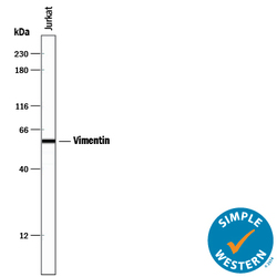
- Detection of Human Vimentin by Western Blot. Western blot shows lysates of Jurkat human acute T cell leukemia cell line and K562 human chronic myelogenous leukemia cell line. PVDF membrane was probed with 2 µg/mL of Rat Anti-Human/Mouse/Rat Vimentin Monoclonal Antibody (Catalog # MAB2105) followed by HRP-conjugated Anti-Rat IgG Secondary Antibody (Catalog # HAF005). A specific band was detected for Vimentin at approximately 55 kDa (as indicated). This experiment was conducted under reducing conditions and using Immunoblot Buffer Group 1.

- Submitted by
- R&D Systems (provider)
- Main image
- Experimental details
- Detection of Human Vimentin by Simple WesternTM. Simple Western lane view shows lysates of Jurkat human acute T cell leukemia cell line, loaded at 0.2 mg/mL. A specific band was detected for Vimentin at approximately 58 kDa (as indicated) using 10 µg/mL of Rat Anti-Human/Mouse/Rat Vimentin Monoclonal Antibody (Catalog # MAB2105) followed by 1:50 dilution of HRP-conjugated Anti-Rat IgG Secondary Antibody (Catalog # HAF005). This experiment was conducted under reducing conditions and using the 12-230 kDa separation system.

- Submitted by
- R&D Systems (provider)
- Main image

- Experimental details
- Detection of Mouse and Rat Vimentin by Western Blot. Western blot shows lysates of MEF mouse embryonic feeder cells, NIH-3T3 mouse embryonic fibroblast cell line, Rat-2 rat embryonic fibroblast cell line, and NR8383 rat alveolar macrophage cell line. PVDF membrane was probed with 1 µg/mL of Rat Anti-Human/Mouse/Rat Vimentin Monoclonal Antibody (Catalog # MAB2105) followed by HRP-conjugated Anti-Rat IgG Secondary Antibody (Catalog # HAF005). A specific band was detected for Vimentin at approximately 55 kDa (as indicated). This experiment was conducted under reducing conditions and using Immunoblot Buffer Group 1.

Supportive validation
- Submitted by
- R&D Systems (provider)
- Main image

- Experimental details
- Vimentin in Mouse Cortical Stem Cells. Vimentin was detected in immersion fixed mouse cortical stem cells using Rat Anti-Human/Mouse/Rat Vimentin Monoclonal Antibody (Catalog # MAB2105) at 10 µg/mL for 3 hours at room temperature. Cells were stained using the NorthernLights™ 557-conjugated Anti-Rat IgG Secondary Antibody (red; Catalog # NL013) and counterstained with DAPI (blue). Specific staining was localized to cytoskeleton. View our protocol for Fluorescent ICC Staining of Cells on Coverslips.

- Submitted by
- R&D Systems (provider)
- Main image

- Experimental details
- Vimentin in Rat Cortical Stem Cells. Vimentin was detected in immersion fixed rat cortical stem cells using Rat Anti-Human/Mouse/Rat Vimentin Monoclonal Antibody (Catalog # MAB2105) at 10 µg/mL for 3 hours at room temperature. Cells were stained using the NorthernLights™ 557-conjugated Anti-Rat IgG Secondary Antibody (red; Catalog # NL013) and counterstained with DAPI (blue). Specific staining was localized to cytoskeleton. View our protocol for Fluorescent ICC Staining of Cells on Coverslips.

- Submitted by
- R&D Systems (provider)
- Main image

- Experimental details
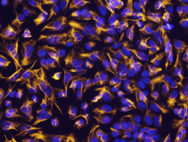
- Vimentin in A549 Human Cell Line. Vimentin was detected in immersion fixed A549 human lung carcinoma cell line using Rat Anti-Human/Mouse/Rat Vimentin Monoclonal Antibody (Catalog # MAB2105) at 10 µg/mL for 3 hours at room temperature. Cells were stained using the NorthernLights™ 493-conjugated Anti-Rat IgG Secondary Antibody (green; Catalog # NL015) and counterstained with DAPI (blue). View our protocol for Fluorescent ICC Staining of Cells on Coverslips.

- Submitted by
- R&D Systems (provider)
- Main image

- Experimental details
- Vimentin in NTera-2 Human Cell Line. Vimentin was detected in immersion fixed NTera-2 human testicular embryonic carcinoma cell line using Rat Anti-Human/Mouse/Rat Vimentin Monoclonal Antibody (Catalog # MAB2105) at 10 µg/mL for 3 hours at room temperature. Cells were stained using the NorthernLights™ 557-conjugated Anti-Rat IgG Secondary Antibody (yellow; Catalog # NL013) and counterstained with DAPI (blue). View our protocol for Fluorescent ICC Staining of Cells on Coverslips.
Supportive validation
- Submitted by
- R&D Systems (provider)
- Main image

- Experimental details
- Vimentin in Human Tonsil. Vimentin was detected in immersion fixed paraffin-embedded sections of human tonsil using Rat Anti-Human/Mouse/Rat Vimentin Monoclonal Antibody (Catalog # MAB2105) at 0.5 µg/mL for 1 hour at room temperature followed by incubation with the Anti-Rat IgG VisUCyte™ HRP Polymer Antibody (Catalog # VC005). Tissue was stained using DAB (brown) and counterstained with hematoxylin (blue). Specific staining was localized to cytoplasm. View our protocol for IHC Staining with VisUCyte HRP Polymer Detection Reagents.

Supportive validation
- Submitted by
- R&D Systems (provider)
- Main image
- Experimental details
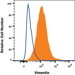
- Detection of Vimentin in A172 Human Cell Line by Flow Cytometry. A172 human glioblastoma cell line was stained with Rat Anti-Human/Mouse/Rat Vimentin Monoclonal Antibody (Catalog # MAB2105, filled histogram) or isotype control antibody (Catalog # MAB006, open histogram) followed by anti-Rat IgG PE-conjugated secondary antibody (Catalog # F0105B). To facilitate intracellular staining, cells were fixed with Flow Cytometry Fixation Buffer (Catalog # FC004) and permeabilized with Flow Cytometry Permeabilization/Wash Buffer I (Catalog # FC005). View our protocol for Staining Intracellular Molecules.
