 Explore
 Validate
 Learn
Learn Western blot
Antibody data
- Antibody Data
- Antigen structure
- References [1]
- Comments [0]
- Validations
- Western blot [1]
- Immunocytochemistry [2]
- Immunohistochemistry [6]
Submit
Validation data
Reference
Comment
Report error
- Product number
- TA506403 - Provider product page

- Provider
- OriGene
- Proper citation
- OriGene Cat#TA506403, RRID:AB_2623760
- Product name
- CDH3 (P cadherin) mouse monoclonal antibody, clone OTI2D5 (formerly 2D5)
- Antibody type
- Monoclonal
- Description
- CDH3 (P cadherin) mouse monoclonal antibody, clone OTI2D5 (formerly 2D5)
- Host
- Mouse
- Conjugate
- Unconjugated
- Epitope
- CDH3
- Isotype
- IgG
- Antibody clone number
- OTI2D5
- Vial size
- 100 µl
- Concentration
- 1 mg/ml
Submitted references Exclusion from spheroid formation identifies loss of essential cell-cell adhesion molecules in colon cancer cells.
Stadler M, Scherzer M, Walter S, Holzner S, Pudelko K, Riedl A, Unger C, Kramer N, Weil B, Neesen J, Hengstschläger M, Dolznig H
Scientific reports 2018 Jan 18;8(1):1151
Scientific reports 2018 Jan 18;8(1):1151
No comments: Submit comment
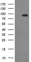
Supportive validation
- Submitted by
- OriGene (provider)
- Main image

- Experimental details
- HEK293T cells were transfected with the pCMV6-ENTRY control (Left lane) or pCMV6-ENTRY CDH3 (RC207346, Right lane) cDNA for 48 hrs and lysed. Equivalent amounts of cell lysates (5 ug per lane) were separated by SDS-PAGE and immunoblotted with anti-CDH3.
- Validation comment
- WB
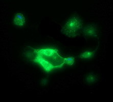
Supportive validation
- Submitted by
- OriGene (provider)
- Main image

- Experimental details
- Anti-CDH3 mouse monoclonal antibody (TA506403) immunofluorescent staining of COS7 cells transiently transfected by pCMV6-ENTRY CDH3(RC207346).
- Validation comment
- IF
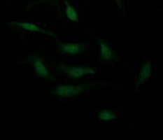
- Submitted by
- OriGene (provider)
- Main image

- Experimental details
- Immunofluorescent staining of HeLa cells using anti-CDH3 mouse monoclonal antibody (TA506403).
- Validation comment
- IF
Supportive validation
- Submitted by
- OriGene (provider)
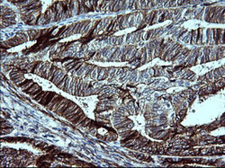
- Main image
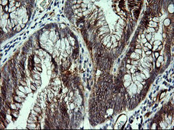
- Experimental details
- Immunohistochemical staining of paraffin-embedded Adenocarcinoma of Human colon tissue using anti-CDH3 mouse monoclonal antibody. (Heat-induced epitope retrieval by 10mM citric buffer, pH6.0, 120C for 3min, TA506403)
- Validation comment
- IHC

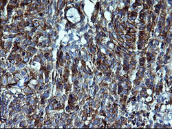
- Submitted by
- OriGene (provider)
- Main image
- Experimental details
- Immunohistochemical staining of paraffin-embedded Adenocarcinoma of Human endometrium tissue using anti-CDH3 mouse monoclonal antibody. (Heat-induced epitope retrieval by 10mM citric buffer, pH6.0, 120C for 3min, TA506403)
- Validation comment
- IHC

- Submitted by
- OriGene (provider)
- Main image
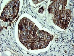
- Experimental details
- Immunohistochemical staining of paraffin-embedded Adenocarcinoma of Human breast tissue using anti-CDH3 mouse monoclonal antibody. (Heat-induced epitope retrieval by 10mM citric buffer, pH6.0, 120C for 3min, TA506403)
- Validation comment
- IHC

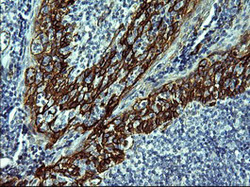
- Submitted by
- OriGene (provider)
- Main image
- Experimental details
- Immunohistochemical staining of paraffin-embedded Carcinoma of Human lung tissue using anti-CDH3 mouse monoclonal antibody. (Heat-induced epitope retrieval by 10mM citric buffer, pH6.0, 120C for 3min, TA506403)
- Validation comment
- IHC

- Submitted by
- OriGene (provider)
- Main image

- Experimental details
- Immunohistochemical staining of paraffin-embedded Human lymphoma tissue using anti-CDH3 mouse monoclonal antibody. (Heat-induced epitope retrieval by 10mM citric buffer, pH6.0, 120C for 3min, TA506403)
- Validation comment
- IHC

- Submitted by
- OriGene (provider)
- Main image
- Experimental details
- Immunohistochemical staining of paraffin-embedded Human lymph node tissue within the normal limits using anti-CDH3 mouse monoclonal antibody. (Heat-induced epitope retrieval by 10mM citric buffer, pH6.0, 120C for 3min, TA506403)
- Validation comment
- IHC
