Explore
 Validate
 Learn
Learn Immunocytochemistry
 Immunohistochemistry
Antibody data
- Antibody Data
- Antigen structure
- References [2]
- Comments [0]
- Validations
- Immunocytochemistry [2]
- Immunohistochemistry [7]
Submit
Validation data
Reference
Comment
Report error
- Product number
- HPA005653 - Provider product page

- Provider
- Atlas Antibodies
- Proper citation
- Atlas Antibodies Cat#HPA005653, RRID:AB_1079293
- Product name
- Anti-MAFB
- Antibody type
- Polyclonal
- Reactivity
- Human
- Host
- Rabbit
- Conjugate
- Unconjugated
- Antigen sequence
MEYVNDFDLLKFDVKKEPLGRAERPGRPCTRLQPA
GSVSSTPLSTPCSSVPSSPSFSPTEQKTHLEDLYW
MASNYQQMNPEALNLTPEDAVEALIGSHPVPQPLQ
SFDSFRGAHHHHHHHHPH- Isotype
- IgG
- Vial size
- 100 µl
- Storage
- Store at +4°C for short term storage. Long time storage is recommended at -20°C.
Submitted references Pancreatic islet enhancer clusters enriched in type 2 diabetes risk-associated variants
Single cell dissection of early kidney development: multilineage priming.
Pasquali L, Gaulton K, Rodríguez-Seguí S, Mularoni L, Miguel-Escalada I, Akerman İ, Tena J, Morán I, Gómez-Marín C, van de Bunt M, Ponsa-Cobas J, Castro N, Nammo T, Cebola I, García-Hurtado J, Maestro M, Pattou F, Piemonti L, Berney T, Gloyn A, Ravassard P, Skarmeta J, Müller F, McCarthy M, Ferrer J
Nature Genetics 2014 January;46(2):136-143
Nature Genetics 2014 January;46(2):136-143
Single cell dissection of early kidney development: multilineage priming.
Brunskill EW, Park JS, Chung E, Chen F, Magella B, Potter SS
Development (Cambridge, England) 2014 Aug;141(15):3093-101
Development (Cambridge, England) 2014 Aug;141(15):3093-101
No comments: Submit comment
Supportive validation
- Submitted by
- Atlas Antibodies (provider)
- Main image
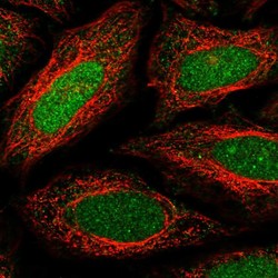
- Experimental details
- Immunofluorescent staining of human cell line SiHa shows localization to nucleus & nucleoli.
- Sample type
- HUMAN

- Submitted by
- Atlas Antibodies (provider)
- Main image

- Experimental details
- Immunofluorescent staining of human cell line hTCEpi shows localization to nucleus & the Golgi apparatus.
- Sample type
- HUMAN

Enhanced validation
Supportive validation
- Submitted by
- Atlas Antibodies (provider)
- Enhanced method
- Orthogonal validation
- Main image

- Experimental details
- Immunohistochemistry analysis in human parathyroid gland and liver tissues using HPA005653 antibody. Corresponding MAFB RNA-seq data are presented for the same tissues.
- Sample type
- HUMAN

Supportive validation
- Submitted by
- Atlas Antibodies (provider)
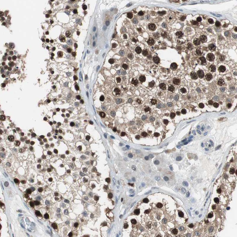
- Main image

- Experimental details
- Immunohistochemical staining of human testis shows strong nuclear positivity in cells of seminiferus ducts.
- Submitted by
- Atlas Antibodies (provider)
- Main image

- Experimental details
- Immunohistochemical staining of human parathyroid gland shows moderate to strong nuclear positivity in glandular cells.
- Sample type
- HUMAN

- Submitted by
- Atlas Antibodies (provider)
- Main image

- Experimental details
- Immunohistochemical staining of human kidney shows moderate to strong nuclear positivity in podocytes.
- Sample type
- HUMAN

- Submitted by
- Atlas Antibodies (provider)
- Main image
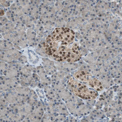
- Experimental details
- Immunohistochemical staining of human pancreas shows moderate to strong nuclear positivity in islets of Langerhans.
- Sample type
- HUMAN

- Submitted by
- Atlas Antibodies (provider)
- Main image

- Experimental details
- Immunohistochemical staining of human lung shows moderate to strong nuclear positivity in macrophages.
- Sample type
- HUMAN

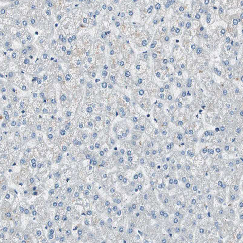
- Submitted by
- Atlas Antibodies (provider)
- Main image

- Experimental details
- Immunohistochemical staining of human liver shows no positivity in hepatocytes as expected.
- Sample type
- HUMAN