Explore
 Validate
 Learn
Learn Western blot
Antibody data
- Antibody Data
- Antigen structure
- References [4]
- Comments [0]
- Validations
- Western blot [2]
- Immunohistochemistry [2]
- Flow cytometry [2]
Submit
Validation data
Reference
Comment
Report error
- Product number
- NBP2-00880 - Provider product page

- Provider
- Novus Biologicals
- Product name
- Mouse Monoclonal RGS5 Antibody
- Antibody type
- Monoclonal
- Description
- Affinity purified.
- Reactivity
- Human
- Host
- Mouse
- Isotype
- IgG
- Vial size
- 0.1 ml
- Concentration
- 1 mg/ml
- Storage
- Store at -20C. Avoid freeze-thaw cycles.
Submitted references Enrichment of CD146+ Adipose-Derived Stem Cells in Combination with Articular Cartilage Extracellular Matrix Scaffold Promotes Cartilage Regeneration.
Programmed death-ligand 1 expression is an unfavorable prognostic factor of hepatocellular carcinoma after archiving sustained virologic response for hepatitis C virus infection.
Expression and role of regulator of G-protein signaling 5 in squamous cell carcinoma of the tongue.
Regulator of G-protein signaling 5 enhances portal vein invasion in hepatocellular carcinoma.
Li X, Guo W, Zha K, Jing X, Wang M, Zhang Y, Hao C, Gao S, Chen M, Yuan Z, Wang Z, Zhang X, Shen S, Li H, Zhang B, Xian H, Zhang Y, Sui X, Qin L, Peng J, Liu S, Lu S, Guo Q
Theranostics 2019;9(17):5105-5121
Theranostics 2019;9(17):5105-5121
Programmed death-ligand 1 expression is an unfavorable prognostic factor of hepatocellular carcinoma after archiving sustained virologic response for hepatitis C virus infection.
Kondo R, Akiba J, Ogasawara S, Nakashima O, Naito Y, Kusano H, Mihara Y, Tanigawa M, Yano H
Oncology letters 2019 Aug;18(2):1458-1466
Oncology letters 2019 Aug;18(2):1458-1466
Expression and role of regulator of G-protein signaling 5 in squamous cell carcinoma of the tongue.
Abe Y, Ogasawara S, Akiba J, Naito Y, Kondo R, Nakamura K, Kusukawa J, Yano H
Clinical and experimental dental research 2019 Apr;5(2):160-169
Clinical and experimental dental research 2019 Apr;5(2):160-169
Regulator of G-protein signaling 5 enhances portal vein invasion in hepatocellular carcinoma.
Umeno Y, Ogasawara S, Akiba J, Hattori S, Kusano H, Nakashima O, Koga H, Torimura T, Yamakawa R, Yano H
Oncology letters 2018 Feb;15(2):1763-1770
Oncology letters 2018 Feb;15(2):1763-1770
No comments: Submit comment
Supportive validation
- Submitted by
- Novus Biologicals (provider)
- Main image
- Experimental details
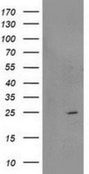
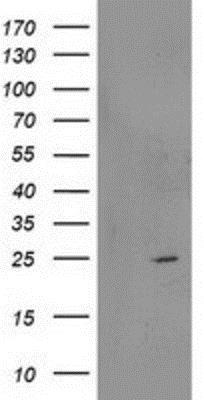
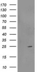
- Western Blot: RGS5 Antibody (1C1) [NBP2-00880] - HEK293T cells were transfected with the pCMV6-ENTRY control (Left lane) or pCMV6-ENTRY RGS5 (Right lane) cDNA for 48 hrs and lysed. Equivalent amounts of cell lysates (5 ug per lane) were separated by SDS-PAGE and immunoblotted with anti-RGS5.
- Submitted by
- Novus Biologicals (provider)
- Main image
- Experimental details
- Western Blot: RGS5 Antibody (OTI1C1) [NBP2-00880] - HEK293T cells were transfected with the pCMV6-ENTRY control (Left lane) or pCMV6-ENTRY RGS5 (Right lane) cDNA for 48 hrs and lysed. Equivalent amounts of cell lysates (5 ug per lane) were separated by SDS-PAGE and immunoblotted with anti-RGS5.

Supportive validation
- Submitted by
- Novus Biologicals (provider)
- Main image
- Experimental details
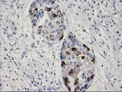
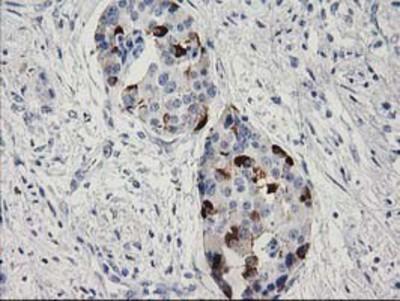
- Immunohistochemistry-Paraffin: RGS5 Antibody (1C1) [NBP2-00880] - Staining of paraffin-embedded Carcinoma of Human pancreas tissue using anti-RGS5 mouse monoclonal antibody.
- Submitted by
- Novus Biologicals (provider)
- Main image
- Experimental details
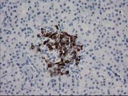
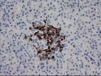
- Immunohistochemistry-Paraffin: RGS5 Antibody (OTI1C1) [NBP2-00880] - Staining of paraffin-embedded Human pancreas tissue within the normal limits using anti-RGS5 mouse monoclonal antibody. (Heat-induced epitope retrieval by 10mM citric buffer, pH6.0, 100 degrees C for 10min)
Supportive validation
- Submitted by
- Novus Biologicals (provider)
- Main image

- Experimental details
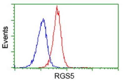
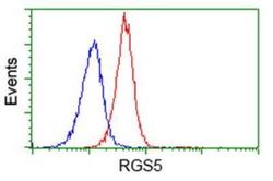
- Flow Cytometry: RGS5 Antibody (OTI1C1) [NBP2-00880] - HEK293T cells transfected with either overexpress plasmid (Red) or empty vector control plasmid (Blue) were immunostained by anti-RGS5 antibody and then analyzed by flow cytometry.
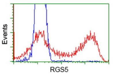
- Submitted by
- Novus Biologicals (provider)
- Main image
- Experimental details
- Flow Cytometry: RGS5 Antibody (OTI1C1) [NBP2-00880] - Flow cytometric Analysis of Jurkat cells, using anti-RGS5 antibody, (Red), compared to a nonspecific negative control antibody, (Blue).