Explore
 Validate
 Learn
Learn Western blot
Antibody data
- Antibody Data
- Antigen structure
- References [0]
- Comments [0]
- Validations
- Western blot [1]
- Flow cytometry [2]
Submit
Validation data
Reference
Comment
Report error
- Product number
- 10-1031-25 - Provider product page

- Provider
- ABEOMICS Inc.
- Product name
- Anti-Human NOXA Antibody
- Antibody type
- Monoclonal
- Description
- Noxa is a BH3-only pro-apoptotic protein that mediates apoptosis by specifically inhibiting the anti-apoptotic Bcl-2 family member Mcl-1. It consists of 54 amino acids with one BH3 domain. The gene for NOXA is a target of p53. In response to cellular stress Noxa is induced by p53 and mediates p53-induced apoptosis. But more recently, it has been shown that NOXA can also be induced independently of p53 by other transcription factors such as p73 and E2F1. Noxa is also found to be induced by hypoxia-inducible factor (HIF-1) and mediates HIF-1 induced hypoxic cell death. Noxa is commonly upregulated in melanomas, and is associated with melanoma development and progression. Increase in Noxa expression is driven by oncogenic activation of MEK/ERK signaling through the transcription factor CREB (cAMP responsive element binding protein).
- Reactivity
- Human
- Host
- Mouse
- Conjugate
- Unconjugated
- Antigen sequence
Full length recombinant protein of
NOXA was used as the immunogen for
this antibody.- Isotype
- IgG
- Antibody clone number
- ABM16G6
- Vial size
- 100 µg
- Concentration
- 0.5 mg/ml
- Storage
- Store the antibody at 4°C, stable for 6 months. For long-term storage, store at -20°C. Avoid repeat freez thawing
No comments: Submit comment
Supportive validation
- Submitted by
- ABEOMICS Inc. (provider)
- Main image
- Experimental details
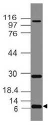
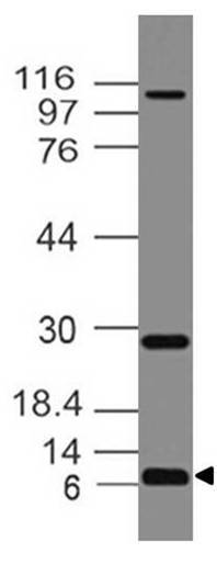
- Western blot analysis of NOXA. Anti- NOXA antibody (Clone: ABM16G6) was used at 4 µg/ml on HCT-116 lysates.
- Protocol
- Protocol
Supportive validation
- Submitted by
- ABEOMICS Inc. (provider)
- Main image
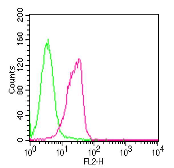
- Experimental details
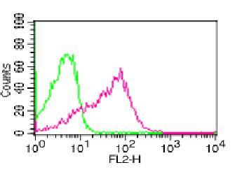
- FLOW analysis of NOXA. Intercellular staining of Jurkat cells. Green represents mouse IgG1 Isotype control (ABEOMICS). Red represents Anti-NOXA antibody. 0.5 ug of antibody was used. Goat anti-mouse PE was used as secondary antibody.
- Protocol
- Protocol
- Submitted by
- ABEOMICS Inc. (provider)
- Main image
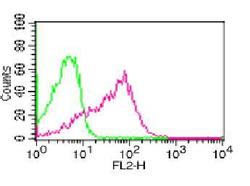
- Experimental details
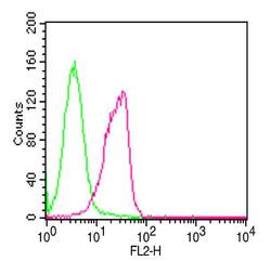
- FLOW analysis of NOXA. Intercellular staining of PBMC. Green represents mouse IgG1 Isotype control (ABEOMICS). Red represents Anti-NOXA antibody. 0.5 ug of antibody was used. Goat anti-mouse PE was used as secondary antibody.
- Protocol
- Protocol