Explore
 Validate
 Learn
Learn Western blot
Antibody data
- Antibody Data
- Antigen structure
- References [1]
- Comments [0]
- Validations
- Western blot [3]
- Immunocytochemistry [2]
- Immunohistochemistry [2]
- Other assay [3]
Submit
Validation data
Reference
Comment
Report error
- Product number
- PA5-19977 - Provider product page

- Provider
- Invitrogen Antibodies
- Product name
- NOXA Polyclonal Antibody
- Antibody type
- Polyclonal
- Antigen
- Synthetic peptide
- Reactivity
- Human, Mouse, Rat
- Host
- Rabbit
- Isotype
- IgG
- Vial size
- 100 µg
- Concentration
- 1 mg/mL
- Storage
- Maintain refrigerated at 2-8°C for up to 3 months. For long term storage store at -20°C
Submitted references Pro-death signaling of cytoprotective heat shock factor 1: upregulation of NOXA leading to apoptosis in heat-sensitive cells.
Janus P, Toma-Jonik A, Vydra N, Mrowiec K, Korfanty J, Chadalski M, Widłak P, Dudek K, Paszek A, Rusin M, Polańska J, Widłak W
Cell death and differentiation 2020 Jul;27(7):2280-2292
Cell death and differentiation 2020 Jul;27(7):2280-2292
No comments: Submit comment
Supportive validation
- Submitted by
- Invitrogen Antibodies (provider)
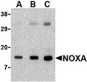
- Main image

- Experimental details
- Western blot analysis of human stomach tissue lysate using a Noxa polyclonal antibody (Product # PA5-19977) at (A) 0.5, (B) 1 and (C) 2 µg/mL.

- Submitted by
- Invitrogen Antibodies (provider)
- Main image

- Experimental details
- Western Blot analysis of Noxa in human stomach tissue lysate with NOXA Polyclonal Antibody (Product # PA5-19977) at (A) 0.5, (B) 1 and (C) 2 µg/mL.
- Submitted by
- Invitrogen Antibodies (provider)
- Main image

- Experimental details
- Western Blot analysis of Noxa in human stomach tissue lysate with NOXA Polyclonal Antibody (Product # PA5-19977) at (A) 0.5, (B) 1 and (C) 2 µg/mL.

Supportive validation
- Submitted by
- Invitrogen Antibodies (provider)
- Main image
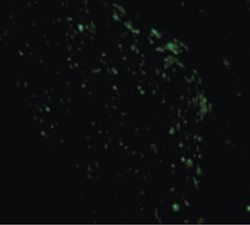
- Experimental details
- Immunofluorescent analysis of human stomach cells using a Noxa polyclonal antibody (Product # PA5-19977) at a 10 µg/mL dilution.
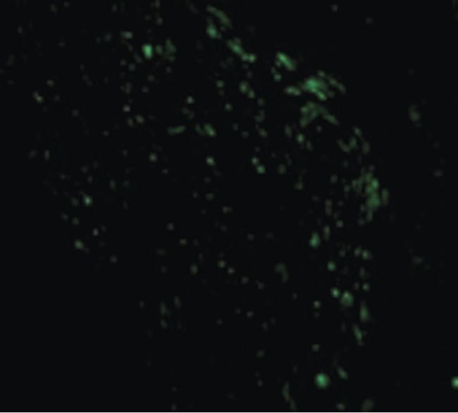
- Submitted by
- Invitrogen Antibodies (provider)
- Main image

- Experimental details
- Immunofluorescence of Noxa in Human Stomach cells with NOXA Polyclonal Antibody (Product # PA5-19977) at 10 µg/mL.

Supportive validation
- Submitted by
- Invitrogen Antibodies (provider)
- Main image

- Experimental details
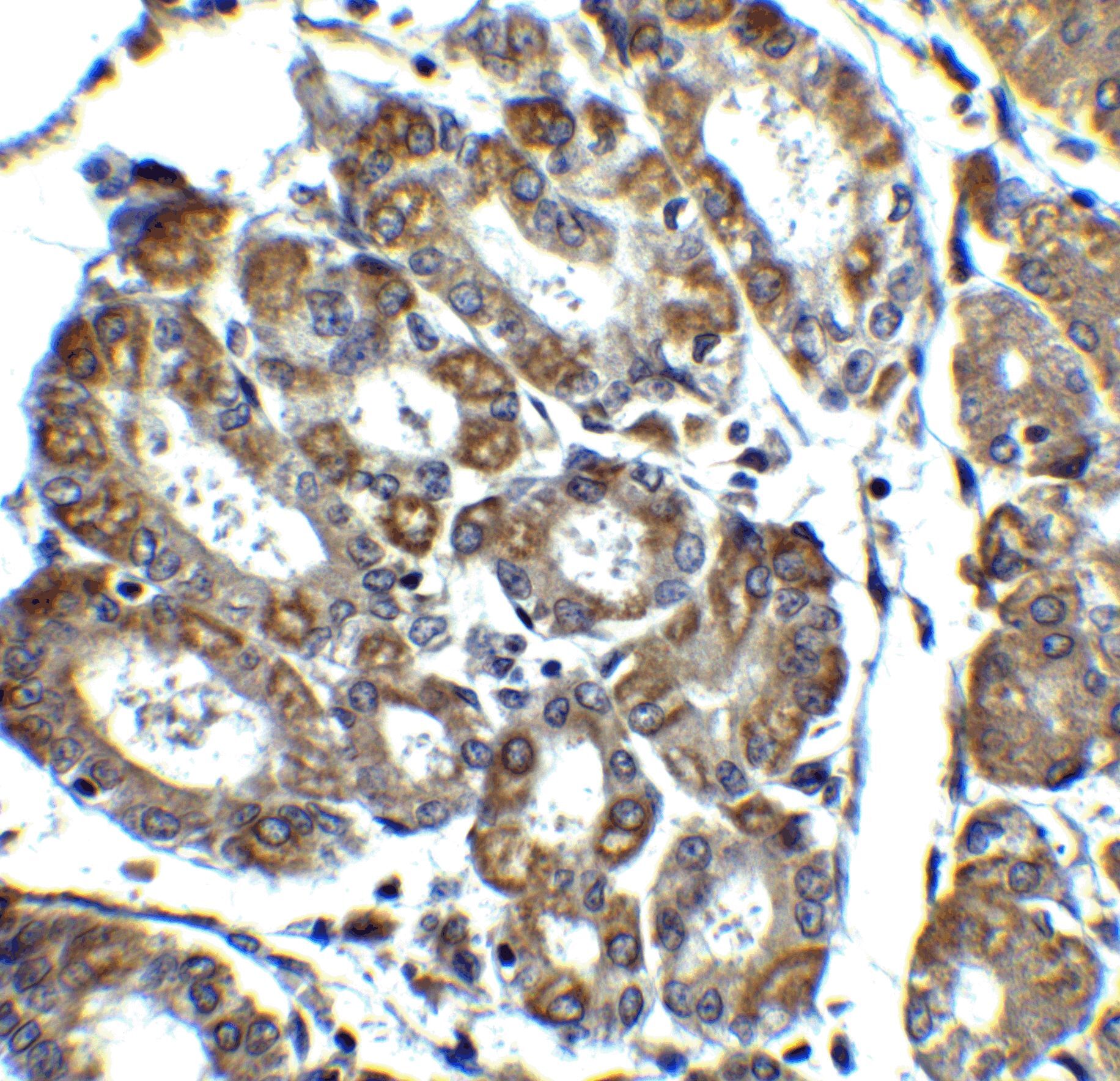
- Immunohistochemistry of Noxa in human stomach tissue with NOXA Polyclonal Antibody (Product # PA5-19977) at 1 µg/mL.

- Submitted by
- Invitrogen Antibodies (provider)
- Main image

- Experimental details
- Immunohistochemistry of NOXA in human stomach tissue with NOXA Polyclonal Antibody (Product # PA5-19977) at 2.5 µg/mL.
Supportive validation
- Submitted by
- Invitrogen Antibodies (provider)
- Main image
- Experimental details
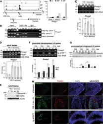
- Fig. 1 Heat shock-induced HSF1 binding in the introns of the Pmaip1 gene correlates with upregulation of its expression and enhanced apoptosis in mouse spermatogenic cells. a Chromatin binding of HSF1 assessed by ChIP-Seq in isolated spermatocytes. Organization of mouse and human genes is shown below peaks of ChIP-Seq tags: bars--exons (darker bars--coding regions), lines--introns; corresponding start and stop codons are linked by light-gray dashed or solid lines, respectively; the positions of HSE or HSE-like motifs are indicated by the closed and open arrows, respectively. Right panel shows the magnitude of HSF1 binding in intronic HSE of the Pmaip1 gene in comparison to Hsph1 promoter based on data from ChIP-Seq extracted from GSE56735. b HSF1 binding in Pmaip1 introns analyzed by ChIP-PCR in isolated spermatocytes. Binding to the Hsph1 promoter is shown as a positive control. C control, physiological temperature of testes (32 degC); 38deg and 43deg, heat shock at 38 or 43 degC, respectively; M marker; - +, negative and positive PCR controls. c Induction of Pmaip1 transcription assayed by RT-PCR and RT-qPCR in isolated spermatocytes after heat shock in vitro at 43 degC and d in testes of mice after heat shock in vivo. 18S rRNA and Hspa1 were used as transcript level controls for loading and the heat shock response, respectively; C control, HS heat shock. e Accumulation of PMAIP1 protein after heat shock in vivo in mouse testes demonstrated by western blot. ACTB and HSPA1 w

- Submitted by
- Invitrogen Antibodies (provider)
- Main image

- Experimental details
- Fig. 2 HSF1 and HSEs present in the introns of Pmaip1 gene are essential for its activation after heat shock. a Heat shock-induced HSF1 binding in introns of Pmaip1 analyzed by ChIP-qPCR in wild-type (WT) HECa10 cells and the clone with hemideletion (1/2HSE) of the perfect HSE in the second intron. b RT-qPCR assays of Pmaip1 and Hspa1a genes transcript levels after heat shock (HS) in cells described in panel a . Fold changes in reference to untreated cells are shown. c PMAIP1 level in the same cells analyzed by western blot. CPT treatment served as positive control for PMAIP1 upregulation. ACTB is shown as a control for loading. Lower panel shows the representative results of densitometric analyses of western blots; * p < 0.05. d Relative luciferase activity in the human 1205Lu cells stably expressing constitutively active HSF1 (aHSF1) in relation to control cells with the empty vector (Neo). Cells were transiently transfected with: the pGL3-Promoter vector (a), its derivatives with the part of the second intron of the mouse Pmaip1 gene acting as an enhancer, containing either wild-type (a1) or mutated HSE (a2), and the vector with the HSPA7 promoter (b) used as a positive control. Sequences of wild-type HSE from the second intron of mouse Pmaip1 (nucleotides 93-112 downstream from the exon2/intron2 boundary) and mutated HSE (mutHSE) are shown above the graph. Hats indicate the most essential G and C nucleotides in the HSE sequence. Presented are mean values and +- SD from th

- Submitted by
- Invitrogen Antibodies (provider)
- Main image
- Experimental details
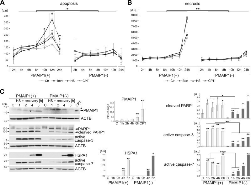
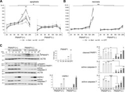
- Fig. 4 PMAIP1 deficiency reduces heat-induced death and delays caspase-3 and caspase-7 activation. a Apoptosis and b necrosis of PMAIP1(+) and PMAIP1(-) HECa10 cells monitored between 2 and 24 h after heat shock or during bortezomib (Bort) or camptothecin (CPT) treatments. Shown are mean values +- SD from one (representative) of three independent experiments; the statistically significant difference between treated and untreated samples or PMAIP1(+) and PMAIP1(-) samples is marked with an asterisk (* p < 0.05, ** p < 0.001). c PMAIP1(+) and PMAIP1(-) cells were heat-shocked for 1 h at 43 degC and protein extracts were analyzed by western blot up to 6 h of recovery. ACTB was used as a loading control. C untreated cells, CPT camptothecin treatment of wild-type HECa10 cells for 6 h (positive control for PMAIP1 induction); unspecific protein band recognized by anti-PMAIP1 Ab is marked with an asterisk. The graphs show the results of densitometric analyses from three independent experiments; * p < 0.05, ** p < 0.001.