 Explore
 Validate
 Learn
Learn Western blot
Antibody data
- Antibody Data
- Antigen structure
- References [7]
- Comments [0]
- Validations
- Western blot [2]
- Immunohistochemistry [1]
Submit
Validation data
Reference
Comment
Report error
- Product number
- AF3150 - Provider product page

- Provider
- Novus Biologicals
- Product name
- Goat Polyclonal FABP4/A-FABP Antibody
- Antibody type
- Polyclonal
- Description
- Antigen Affinity-purified. Detects human FABP4/A-FABP in direct ELISAs and Western blots. In direct ELISAs, approximately 60% cross-reactivity with recombinant mouse (rm) FABP4 is observed, approximately 30% cross-reactivity with recombinant human (rh) FABP3 is observed, and less than 5% cross-reactivity with rhFABP1, -2, -5, -6, -7, -8, -9, and rmFABP9 is observed.
- Reactivity
- Human
- Host
- Goat
- Conjugate
- Unconjugated
- Isotype
- IgG
- Vial size
- 100 ug
- Concentration
- LYOPH
- Storage
- Use a manual defrost freezer and avoid repeated freeze-thaw cycles. 12 months from date of receipt, -20 to -70 degreesC as supplied. 1 month, 2 to 8 degreesC under sterile conditions after reconstitution. 6 months, -20 to -70 degreesC under sterile conditions after reconstitution.
Submitted references Patient Adipose Stem Cell-Derived Adipocytes Reveal Genetic Variation that Predicts Antidiabetic Drug Response.
Firemaster® 550 and its components isopropylated triphenyl phosphate and triphenyl phosphate enhance adipogenesis and transcriptional activity of peroxisome proliferator activated receptor (Pparγ) on the adipocyte protein 2 (aP2) promoter.
Identification of Multipotent Stem Cells in Human Brain Tissue Following Stroke.
Transcriptome sequencing wide functional analysis of human mesenchymal stem cells in response to TLR4 ligand.
Identification of a cell-of-origin for fibroblasts comprising the fibrotic reticulum in idiopathic pulmonary fibrosis.
Derivation and expansion using only small molecules of human neural progenitors for neurodegenerative disease modeling.
A defined glycosaminoglycan-binding substratum for human pluripotent stem cells.
Hu W, Jiang C, Guan D, Dierickx P, Zhang R, Moscati A, Nadkarni GN, Steger DJ, Loos RJF, Hu C, Jia W, Soccio RE, Lazar MA
Cell stem cell 2019 Feb 7;24(2):299-308.e6
Cell stem cell 2019 Feb 7;24(2):299-308.e6
Firemaster® 550 and its components isopropylated triphenyl phosphate and triphenyl phosphate enhance adipogenesis and transcriptional activity of peroxisome proliferator activated receptor (Pparγ) on the adipocyte protein 2 (aP2) promoter.
Tung EWY, Ahmed S, Peshdary V, Atlas E
PloS one 2017;12(4):e0175855
PloS one 2017;12(4):e0175855
Identification of Multipotent Stem Cells in Human Brain Tissue Following Stroke.
Tatebayashi K, Tanaka Y, Nakano-Doi A, Sakuma R, Kamachi S, Shirakawa M, Uchida K, Kageyama H, Takagi T, Yoshimura S, Matsuyama T, Nakagomi T
Stem cells and development 2017 Jun 1;26(11):787-797
Stem cells and development 2017 Jun 1;26(11):787-797
Transcriptome sequencing wide functional analysis of human mesenchymal stem cells in response to TLR4 ligand.
Kim SH, Das A, Chai JC, Binas B, Choi MR, Park KS, Lee YS, Jung KH, Chai YG
Scientific reports 2016 Jul 22;6:30311
Scientific reports 2016 Jul 22;6:30311
Identification of a cell-of-origin for fibroblasts comprising the fibrotic reticulum in idiopathic pulmonary fibrosis.
Xia H, Bodempudi V, Benyumov A, Hergert P, Tank D, Herrera J, Braziunas J, Larsson O, Parker M, Rossi D, Smith K, Peterson M, Limper A, Jessurun J, Connett J, Ingbar D, Phan S, Bitterman PB, Henke CA
The American journal of pathology 2014 May;184(5):1369-83
The American journal of pathology 2014 May;184(5):1369-83
Derivation and expansion using only small molecules of human neural progenitors for neurodegenerative disease modeling.
Reinhardt P, Glatza M, Hemmer K, Tsytsyura Y, Thiel CS, Höing S, Moritz S, Parga JA, Wagner L, Bruder JM, Wu G, Schmid B, Röpke A, Klingauf J, Schwamborn JC, Gasser T, Schöler HR, Sterneckert J
PloS one 2013;8(3):e59252
PloS one 2013;8(3):e59252
A defined glycosaminoglycan-binding substratum for human pluripotent stem cells.
Klim JR, Li L, Wrighton PJ, Piekarczyk MS, Kiessling LL
Nature methods 2010 Dec;7(12):989-94
Nature methods 2010 Dec;7(12):989-94
No comments: Submit comment
Supportive validation
- Submitted by
- Novus Biologicals (provider)
- Main image

- Experimental details
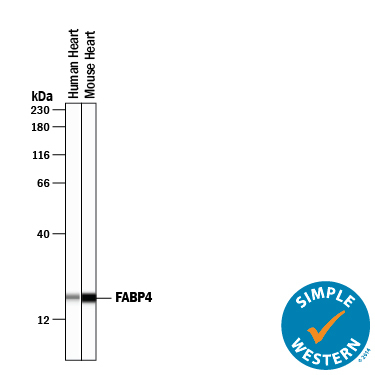
- Detection of Human and Mouse FABP4/A-FABP by Simple WesternTM. Simple Western lane view shows lysates of human heart tissue and mouse heart tissue, loaded at 0.2 mg/mL. A specific band was detected for FABP4/A-FABP at approximately 19 kDa (as indicated) using 10 µg/mL of Goat Anti-Human FABP4/A-FABP Antigen Affinity-purified Polyclonal Antibody (Catalog # AF3150) followed by 1:50 dilution of HRP-conjugated Anti-Goat IgG Secondary Antibody (Catalog # HAF109). This experiment was conducted under reducing conditions and using the 12-230 kDa separation system.
- Submitted by
- Novus Biologicals (provider)
- Main image
- Experimental details
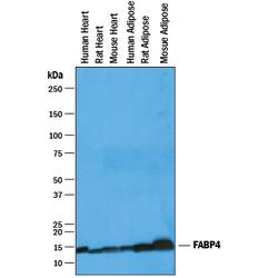
- Detection of Human, Mouse, and Rat FABP4/A-FABP by Western Blot. Western blot shows lysates of human heart tissue, rat heart tissue, mouse heart tissue, human adipose tissue, rat adipose tissue, and mouse adipose tissue. PVDF membrane was probed with 1 µg/mL of Goat Anti-Human FABP4/A-FABP Antigen Affinity-purified Polyclonal Antibody (Catalog # AF3150) followed by HRP-conjugated Anti-Goat IgG Secondary Antibody (Catalog # HAF019). A specific band was detected for FABP4/A-FABP at approximately 14 kDa (as indicated). This experiment was conducted under reducing conditions and using Immunoblot Buffer Group 1.

Supportive validation
- Submitted by
- Novus Biologicals (provider)
- Main image
- Experimental details
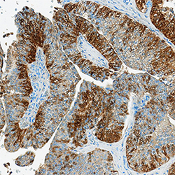
- FABP4/A-FABP in Human Bladder Cancer Tissue. FABP4/A-FABP was detected in immersion fixed paraffin-embedded sections of human bladder cancer tissue using Goat Anti-Human FABP4/A-FABP Antigen Affinity-purified Polyclonal Antibody (Catalog # AF3150) at 3 µg/mL overnight at 4 °C. Tissue was stained using the Anti-Goat HRP-DAB Cell & Tissue Staining Kit (brown; Catalog # CTS008) and counterstained with hematoxylin (blue). Specific staining was localized to cytoplasm. View our protocol for Chromogenic IHC Staining of Paraffin-embedded Tissue Sections.
