 Explore
 Validate
 Learn
Learn Western blot
Antibody data
- Antibody Data
- Antigen structure
- References [4]
- Comments [0]
- Validations
- Western blot [3]
- Immunocytochemistry [2]
- Immunohistochemistry [1]
- Flow cytometry [1]
Submit
Validation data
Reference
Comment
Report error
- Product number
- PA5-16658 - Provider product page

- Provider
- Invitrogen Antibodies
- Product name
- AFP Polyclonal Antibody
- Antibody type
- Polyclonal
- Antigen
- Purifed from natural sources
- Reactivity
- Human, Porcine
- Host
- Rabbit
- Isotype
- IgG
- Vial size
- 500 µL
- Storage
- 4° C
Submitted references Convergence of cMyc and β-catenin on Tcf7l1 enables endoderm specification.
Culture of mouse amniotic fluid-derived cells on irradiated STO feeders results in the generation of primitive endoderm cell lines capable of self-renewal in vitro.
Efficient generation of human iPSCs by a synthetic self-replicative RNA.
Differential proteome and transcriptome analysis of porcine skeletal muscle during development.
Morrison G, Scognamiglio R, Trumpp A, Smith A
The EMBO journal 2016 Feb 1;35(3):356-68
The EMBO journal 2016 Feb 1;35(3):356-68
Culture of mouse amniotic fluid-derived cells on irradiated STO feeders results in the generation of primitive endoderm cell lines capable of self-renewal in vitro.
Babic AM, Jang S, Nicolov E, Voicu H, Luckey CJ
Cells, tissues, organs 2013;198(2):111-26
Cells, tissues, organs 2013;198(2):111-26
Efficient generation of human iPSCs by a synthetic self-replicative RNA.
Yoshioka N, Gros E, Li HR, Kumar S, Deacon DC, Maron C, Muotri AR, Chi NC, Fu XD, Yu BD, Dowdy SF
Cell stem cell 2013 Aug 1;13(2):246-54
Cell stem cell 2013 Aug 1;13(2):246-54
Differential proteome and transcriptome analysis of porcine skeletal muscle during development.
Xu Y, Qian H, Feng X, Xiong Y, Lei M, Ren Z, Zuo B, Xu D, Ma Y, Yuan H
Journal of proteomics 2012 Apr 3;75(7):2093-108
Journal of proteomics 2012 Apr 3;75(7):2093-108
No comments: Submit comment
Supportive validation
- Submitted by
- Invitrogen Antibodies (provider)
- Main image
- Experimental details
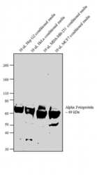
- Western blot analysis was performed on 10 uL conditioned media from Hep G2 (Lane 1), HeLa (Lane 2), MDA-MB-231 (Lane 3) and MCF7 (Lane 4). The blots were probed with Anti-Alpha fetoprotein Rabbit Polyclonal Antibody (Product # PA5-16658, 1:250 dilution) and detected by chemiluminescence using Goat anti-Rabbit IgG (H+L) Superclonal™ Secondary Antibody, HRP conjugate (Product # A27036, 0.4 µg/mL, 1:2500 dilution). A 69 kDa band corresponding to Alpha-fetoprotein was observed across the conditioned media tested. Known quantity of protein samples were electrophoresed using Novex® NuPAGE® 4-12 % Bis-Tris gel (Product # NP0321BOX), XCell SureLock™ Electrophoresis System (Product # EI0002) and Novex® Sharp Pre-Stained Protein Standard (Product # LC5800). Resolved proteins were then transferred onto a nitrocellulose membrane with iBlot® 2 Dry Blotting System (Product # IB21001). The membrane was probed with the relevant primary and secondary Antibody following blocking with 5 % skimmed milk. Chemiluminescent detection was performed using Pierce™ ECL Western Blotting Substrate (Product # 32106).

- Submitted by
- Invitrogen Antibodies (provider)
- Main image

- Experimental details
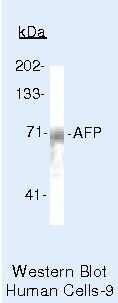
- Western blot of Alpha Fetoprotein using Alpha Fetoprotein Polyclonal Antibody (Product # PA5-16658) on HEP-G-2 Cells.
- Submitted by
- Invitrogen Antibodies (provider)
- Main image
- Experimental details
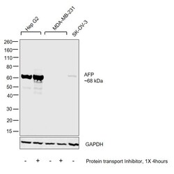
- Western blot was performed using Anti-AFP Polyclonal Antibody (Product # PA5-16658) and a 68 kDa band corresponding to AFP was observed in Hep G2 and SK-OV-3 cells but not in MDA-MB-231 cells which are reported to be negative. Whole cell extracts (30 µg lysate) of Hep G2 (Lane 1), Hep G2 treated with 1X protein transport inhibitor for 4 hours (Product # 00-4980-03) (Lane 2), MDA-MB-231 (Lane 3), MDA-MB-231 treated with 1X protein transport inhibitor for 4 hours (Product # 00-4980-03) (Lane 4) and SK-OV-3 (Lane 5) were electrophoresed using Novex® NuPAGE® 4-12 % Bis-Tris gel (Product # NP0322BOX). Resolved proteins were then transferred onto a nitrocellulose membrane (Product # IB23001) by iBlot® 2 Dry Blotting System (Product # IB21001). The blot was probed with the primary antibody (1:1000 dilution) and detected by chemiluminescence with Goat anti-Rabbit IgG (H+L), Superclonal™ Recombinant Secondary Antibody, HRP (Product # A27036, 1:4000 dilution) using the iBright FL 1000 (Product # A32752). Chemiluminescent detection was performed using Novex® ECL Chemiluminescent Substrate Reagent Kit (Product # WP20005).

Supportive validation
- Submitted by
- Invitrogen Antibodies (provider)
- Main image

- Experimental details
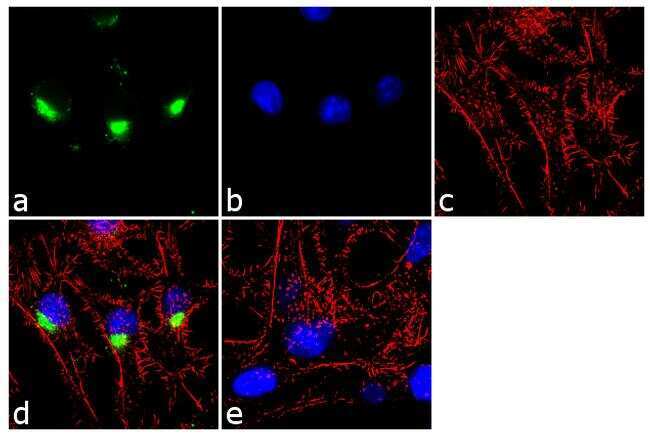
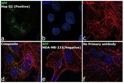
- Immunofluorescence analysis of Alpha Fetoprotein was performed using 70% confluent log phase Hep G2 cells. The cells were fixed with 4% paraformaldehyde for 10 minutes, permeabilized with 0.1% Triton™ X-100 for 10 minutes, and blocked with 1% BSA for 1 hour at room temperature. The cells were labeled with Alpha Fetoprotein Rabbit Polyclonal Antibody (Product # PA5-16658) at 1:250 dilution in 0.1% BSA and incubated for 3 hours at room temperature and then labeled with Goat anti-Rabbit IgG (H+L) Superclonal™ Secondary Antibody, Alexa Fluor® 488 conjugate (Product # A27034) at a dilution of 1:2000 for 45 minutes at room temperature (Panel a: green). Nuclei (Panel b: blue) were stained with SlowFade® Gold Antifade Mountant with DAPI (Product # S36938). F-actin (Panel c: red) was stained with Alexa Fluor® 555 Rhodamine Phalloidin (Product # R415, 1:300). Panel d represents the merged image showing cytoplasmic localization. Panel e shows the no primary antibody control. The images were captured at 60X magnification.
- Submitted by
- Invitrogen Antibodies (provider)
- Main image
- Experimental details
- Immunofluorescence analysis of AFP was performed using 70% confluent log phase Hep G2 and MDA-MB-231 cells. The cells were fixed with 4% paraformaldehyde for 10 minutes, permeabilized with 0.1% Triton™ X-100 for 15 minutes, and blocked with 2% BSA for 1 hour at room temperature. The cells were labeled with AFP Mouse Monoclonal Antibody (Product # PA5-16658) at 1:100 dilution in 0.1% BSA and incubated overnight at 4 degree and then labeled with Donkey anti-Rabbit IgG (H+L) Highly Cross-Adsorbed Secondary Antibody, Alexa Fluor Plus 488 conjugate (Product # A32790) at a dilution of 1:2000 for 45 minutes at room temperature (Panel a: green). Nuclei (Panel b: blue) were stained with ProLong™ Diamond Antifade Mountant with DAPI (Product # P36962). F-actin (Panel c: red) was stained with Rhodamine Phalloidin (Product # R415, 1:300). Panel d represents the composite image showing cytoplasmic and Golgi staining of AFP in Hep G2 cells. Panel e shows MDA-MB-231 cells with no signal. Panel f represents control cells with no primary antibody to assess background. The images were captured at 60X magnification.

Supportive validation
- Submitted by
- Invitrogen Antibodies (provider)
- Main image

- Experimental details
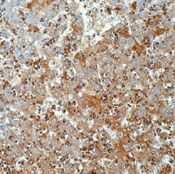
- Formalin-fixed, paraffin-embedded human fetal liver stained with Alpha Fetoprotein antibody using peroxidase-conjugate and DAB chromogen. Note cytoplasmic staining of fetal hepatocytes.
Supportive validation
- Submitted by
- Invitrogen Antibodies (provider)
- Main image
- Experimental details
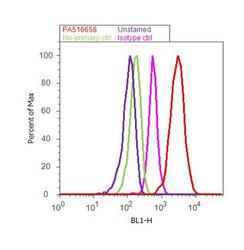
- Flow cytometry analysis of Alpha fetoprotein was done on Hep G2 cells. Cells were fixed with 70% ethanol for 10 minutes, permeabilized with 0.25% Triton™ X-100 for 20 minutes, and blocked with 5% BSA for 30 minutes at room temperature. Cells were labeled with Alpha fetoprotein Rabbit Polyclonal Antibody (Product # PA5-16658, red histogram) or with rabbit isotype control (pink histogram) at 3-5 µg/million cells in 2.5% BSA. After incubation at room temperature for 2 hours, the cells were labeled with Alexa Fluor® 488 Goat Anti-Rabbit Secondary Antibody (Product # A11008) at a dilution of 1:400 for 30 minutes at room temperature. The representative 10,000 cells were acquired and analyzed for each sample using an Attune® Acoustic Focusing Cytometer. The purple histogram represents unstained control cells and the green histogram represents no-primary-antibody control..
