Antibody data
- Antibody Data
- Antigen structure
- References [5]
- Comments [0]
- Validations
- Immunocytochemistry [1]
- Immunohistochemistry [3]
- Flow cytometry [1]
- Other assay [2]
Submit
Validation data
Reference
Comment
Report error
- Product number
- PA5-16918 - Provider product page

- Provider
- Invitrogen Antibodies
- Product name
- CD138 Polyclonal Antibody
- Antibody type
- Polyclonal
- Antigen
- Recombinant full-length protein
- Description
- PA5-16918 targets CD138 in IHC applications and shows reactivity with Human samples.
- Concentration
- 0.046 mg/mL
Submitted references Vascular Inflammation and Dysfunction in Lupus-Prone Mice-IL-6 as Mediator of Disease Initiation.
Globular C1q Receptor (gC1qR/p32/HABP1) Suppresses the Tumor-Inhibiting Role of C1q and Promotes Tumor Proliferation in 1q21-Amplified Multiple Myeloma.
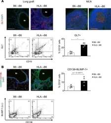
Inhibition of B cell-dependent lymphoid follicle formation prevents lymphocytic bronchiolitis after lung transplantation.
Low levels of monoclonal small B cells in the bone marrow of patients with diffuse large B-cell lymphoma of activated B-cell type but not of germinal center B-cell type.
Proliferation of immature plasma cells in pouchitis mucosa in patients with ulcerative colitis.
Marczynski P, Meineck M, Xia N, Li H, Kraus D, Roth W, Möckel T, Boedecker S, Schwarting A, Weinmann-Menke J
International journal of molecular sciences 2021 Feb 25;22(5)
International journal of molecular sciences 2021 Feb 25;22(5)
Globular C1q Receptor (gC1qR/p32/HABP1) Suppresses the Tumor-Inhibiting Role of C1q and Promotes Tumor Proliferation in 1q21-Amplified Multiple Myeloma.
Xu J, Sun Y, Jiang J, Xu Z, Li J, Xu T, Liu P
Frontiers in immunology 2020;11:1292
Frontiers in immunology 2020;11:1292
Inhibition of B cell-dependent lymphoid follicle formation prevents lymphocytic bronchiolitis after lung transplantation.
Smirnova NF, Conlon TM, Morrone C, Dorfmuller P, Humbert M, Stathopoulos GT, Umkehrer S, Pfeiffer F, Yildirim AÖ, Eickelberg O
JCI insight 2019 Feb 7;4(3)
JCI insight 2019 Feb 7;4(3)
Low levels of monoclonal small B cells in the bone marrow of patients with diffuse large B-cell lymphoma of activated B-cell type but not of germinal center B-cell type.
Tierens AM, Holte H, Warsame A, Ikonomou IM, Wang J, Chan WC, Delabie J
Haematologica 2010 Aug;95(8):1334-41
Haematologica 2010 Aug;95(8):1334-41
Proliferation of immature plasma cells in pouchitis mucosa in patients with ulcerative colitis.
Hirata N, Oshitani N, Kamata N, Sogawa M, Yamagami H, Watanabe K, Watanabe T, Tominaga K, Fujiwara Y, Maeda K, Hirakawa K, Arakawa T
Inflammatory bowel diseases 2008 Aug;14(8):1084-90
Inflammatory bowel diseases 2008 Aug;14(8):1084-90
No comments: Submit comment
Supportive validation
- Submitted by
- Invitrogen Antibodies (provider)
- Main image
- Experimental details
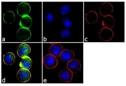
- Immunofluorescence analysis of Syndecan-1 / CD138 was performed using 70% confluent log phase COLO 205 cells. The cells were fixed with 4% paraformaldehyde for 10 minutes, permeabilized with 0.1% Triton™ X-100 for 10 minutes, and blocked with 1% BSA for 1 hour at room temperature. The cells were labeled with Syndecan-1 / CD138 Rabbit Polyclonal Antibody (Product # PA5-16918) at 2µg/mL in 0.1% BSA and incubated for 3 hours at room temperature and then labeled with Goat anti-Rabbit IgG (H+L) Superclonal™ Secondary Antibody, Alexa Fluor® 488 conjµgate (Product # A27034) at a dilution of 1:2000 for 45 minutes at room temperature (Panel a: green). Nuclei (Panel b: blue) were stained with SlowFade® Gold Antifade Mountant with DAPI (Product # S36938). F-actin (Panel c: red) was stained with Alexa Fluor® 555 Rhodamine Phalloidin (Product # R415, 1:300). Panel d represents the merged image showing cytoplasmic and nuclear localization. Panel e shows the no primary antibody control. The images were captured at 60X magnification.
Supportive validation
- Submitted by
- Invitrogen Antibodies (provider)
- Main image
- Experimental details
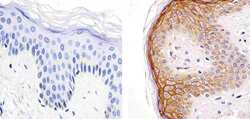
- Immunohistochemistry analysis of Syndecan-1/CD138 showing staining in the cytoplasm and membrane of paraffin-embedded human skin tissue (right) compared to a negative control without primary antibody (left). To expose target proteins, antigen retrieval was performed using 10mM sodium citrate (pH 6.0), microwaved for 8-15 min. Following antigen retrieval, tissues were blocked in 3% H2O2-methanol for 15 min at room temperature, washed with ddH2O and PBS, and then probed with a Syndecan-1/CD138 Rabbit Polyclonal Antibody (Product # PA5-16918) diluted in 3% BSA-PBS at a dilution of 1:20 for 1 hour at 37°C in a humidified chamber. Tissues were washed extensively in PBST and detection was performed using an HRP-conjugated secondary antibody followed by colorimetric detection using a DAB kit. Tissues were counterstained with hematoxylin and dehydrated with ethanol and xylene to prep for mounting.
- Submitted by
- Invitrogen Antibodies (provider)
- Main image
- Experimental details
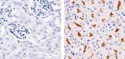
- Immunohistochemistry analysis of Syndecan-1/CD138 showing staining in the membrane of paraffin-embedded mouse kidney tissue (right) compared to a negative control without primary antibody (left). To expose target proteins, antigen retrieval was performed using 10mM sodium citrate (pH 6.0), microwaved for 8-15 min. Following antigen retrieval, tissues were blocked in 3% H2O2-methanol for 15 min at room temperature, washed with ddH2O and PBS, and then probed with a Syndecan-1/CD138 Rabbit Polyclonal Antibody (Product # PA5-16918) diluted in 3% BSA-PBS at a dilution of 1:20 for 1 hour at 37°C in a humidified chamber. Tissues were washed extensively in PBST and detection was performed using an HRP-conjugated secondary antibody followed by colorimetric detection using a DAB kit. Tissues were counterstained with hematoxylin and dehydrated with ethanol and xylene to prep for mounting.
- Submitted by
- Invitrogen Antibodies (provider)
- Main image
- Experimental details
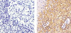
- Immunohistochemistry analysis of Syndecan-1/CD138 showing staining in the cytoplasm and membrane of paraffin-embedded human tonsil tissue (right) compared to a negative control without primary antibody (left). To expose target proteins, antigen retrieval was performed using 10mM sodium citrate (pH 6.0), microwaved for 8-15 min. Following antigen retrieval, tissues were blocked in 3% H2O2-methanol for 15 min at room temperature, washed with ddH2O and PBS, and then probed with a Syndecan-1/CD138 Rabbit Polyclonal Antibody (Product # PA5-16918) diluted in 3% BSA-PBS at a dilution of 1:20 for 1 hour at 37°C in a humidified chamber. Tissues were washed extensively in PBST and detection was performed using an HRP-conjugated secondary antibody followed by colorimetric detection using a DAB kit. Tissues were counterstained with hematoxylin and dehydrated with ethanol and xylene to prep for mounting.
Supportive validation
- Submitted by
- Invitrogen Antibodies (provider)
- Main image
- Experimental details
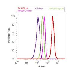
- Flow cytometry analysis of Syndecan-1 / CD138 was done on MCF7 cells. Cells were fixed with 70% ethanol for 10 minutes, permeabilized with 0.25% Triton™ X-100 for 20 minutes, and blocked with 5% BSA for 30 minutes at room temperature. Cells were labeled with Syndecan-1 / CD138 Rabbit Polyclonal Antibody (Product # PA5-16918, red histogram) or with rabbit isotype control (pink histogram) at 3-5 µg/million cells in 2.5% BSA. After incubation at room temperature for 2 hours, the cells were labeled with Alexa Fluor® 488 Goat Anti-Rabbit Secondary Antibody (Product # A11008) at a dilution of 1:400 for 30 minutes at room temperature. The representative 10,000 cells were acquired and analyzed for each sample using an Attune® Acoustic Focusing Cytometer. The purple histogram represents unstained control cells and the green histogram represents no-primary-antibody control..
Supportive validation
- Submitted by
- Invitrogen Antibodies (provider)
- Main image
- Experimental details
- NULL
- Submitted by
- Invitrogen Antibodies (provider)
- Main image
- Experimental details
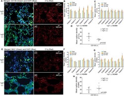
- Figure 2 The amount of C1q deposited around the CD138 + cells in bone marrow (BM) biopsy sections was significantly higher, especially in the 1q21 amplification (Amp1q21) subgroup. (A) Double immunofluorescence labeling on BM biopsy sections: anti-CD138 antibody (green fluorescence) and anti-C1q antibody (red fluorescence). (B) Mean optical density (MOD) of C1q around CD138 + cells and CD138 - cells in the 1q21(+) group. Error bars represent mean +- SD. (C) MOD of C1q around CD138 + cells and CD138 - cells in the 1q21(-) group. Error bars represent mean +- SD. (D) MOD of C1q around CD138 + in comparison with the 1q21(+) and 1q21(-) groups ( p = 0.032). Error bars represent mean +- SD. (E) Double immunofluorescence labeling on BM biopsy sections: anti-CD138 antibody (green fluorescence) and anti-membrane attack complex (anti-MAC) antibody (red fluorescence). (F) MOD of MAC around CD138 + cells and CD138 - cells in the 1q21(+) group. Error bars represent mean +- SD. (G) MOD of MAC around CD138 + cells and CD138 - cells in the 1q21(-) group. Error bars represent mean +- SD. (H) MOD of MAC around CD138 + in comparison with the 1q21(+) and 1q21(-) groups ( p = 0.400). Error bars represent mean +- SD.
 Explore
 Validate
 Learn
Learn Immunocytochemistry