 Explore
 Validate
 Learn
Learn Western blot
 Immunocytochemistry
Antibody data
- Antibody Data
- Antigen structure
- References [12]
- Comments [0]
- Validations
- Western blot [5]
- Immunocytochemistry [2]
- Immunohistochemistry [1]
Submit
Validation data
Reference
Comment
Report error
- Product number
- GTX102156 - Provider product page

- Provider
- GeneTex
- Proper citation
- GeneTex Cat#GTX102156, RRID:AB_1949971
- Product name
- Cofilin 1 antibody
- Antibody type
- Polyclonal
- Reactivity
- Human, Mouse, Rat
- Host
- Rabbit
Submitted references Proteomic analysis of evodiamine-induced cytotoxicity in thyroid cancer cells.
Identification of CRKII, CFL1, CNTN1, NME2, and TKT as Novel and Frequent T-Cell Targets in Human IDH-Mutant Glioma.
Proteomic investigating the cooperative lethal effect of EGFR and MDM2 inhibitors on ovarian carcinoma.
Merkel Cell Polyomavirus Small T Antigen Drives Cell Motility via Rho-GTPase-Induced Filopodium Formation.
Proteomic analysis of honokiol-induced cytotoxicity in thyroid cancer cells.
The putative involvement of actin-binding proteins and cytoskeleton proteins in pathological mechanisms of ketamine cystitis-Revealed by a prospective pilot study using proteomic approaches.
Over-expression of cofilin-1 suppressed growth and invasion of cancer cells is associated with up-regulation of let-7 microRNA.
Nox4-dependent activation of cofilin mediates VSMC reorientation in response to cyclic stretching.
14-3-3ε and ζ regulate neurogenesis and differentiation of neuronal progenitor cells in the developing brain.
A comparative study of primary and recurrent human glioblastoma multiforme using the small animal imaging and molecular expressive profiles.
Hyaluronic acid-dependent protection against alkali-burned human corneal cells.
Platelet-derived growth factor (PDGF) regulates Slingshot phosphatase activity via Nox1-dependent auto-dephosphorylation of serine 834 in vascular smooth muscle cells.
Yu HI, Chou HC, Su YC, Lin LH, Lu CH, Chuang HH, Tsai YT, Liao EC, Wei YS, Yang YT, Lee YR, Chan HL
Journal of pharmaceutical and biomedical analysis 2018 Oct 25;160:344-350
Journal of pharmaceutical and biomedical analysis 2018 Oct 25;160:344-350
Identification of CRKII, CFL1, CNTN1, NME2, and TKT as Novel and Frequent T-Cell Targets in Human IDH-Mutant Glioma.
Dettling S, Stamova S, Warta R, Schnölzer M, Rapp C, Rathinasamy A, Reuss D, Pocha K, Roesch S, Jungk C, Warnken U, Eckstein V, Grabe N, Schramm C, Weigand MA, von Deimling A, Unterberg A, Beckhove P, Herold-Mende C
Clinical cancer research : an official journal of the American Association for Cancer Research 2018 Jun 15;24(12):2951-2962
Clinical cancer research : an official journal of the American Association for Cancer Research 2018 Jun 15;24(12):2951-2962
Proteomic investigating the cooperative lethal effect of EGFR and MDM2 inhibitors on ovarian carcinoma.
Chang SJ, Liao EC, Yeo HY, Kuo WH, Chen HY, Tsai YT, Wei YS, Chen YJ, Wang YS, Li JM, Shih CC, Chan CH, Lai ZY, Chou HC, Chuang YJ, Chan HL
Archives of biochemistry and biophysics 2018 Jun 1;647:10-32
Archives of biochemistry and biophysics 2018 Jun 1;647:10-32
Merkel Cell Polyomavirus Small T Antigen Drives Cell Motility via Rho-GTPase-Induced Filopodium Formation.
Stakaitytė G, Nwogu N, Dobson SJ, Knight LM, Wasson CW, Salguero FJ, Blackbourn DJ, Blair GE, Mankouri J, Macdonald A, Whitehouse A
Journal of virology 2018 Jan 15;92(2)
Journal of virology 2018 Jan 15;92(2)
Proteomic analysis of honokiol-induced cytotoxicity in thyroid cancer cells.
Chou HC, Lu CH, Su YC, Lin LH, Yu HI, Chuang HH, Tsai YT, Liao EC, Wei YS, Yang YT, Chien YA, Yu XR, Lee YR, Chan HL
Life sciences 2018 Aug 15;207:184-204
Life sciences 2018 Aug 15;207:184-204
The putative involvement of actin-binding proteins and cytoskeleton proteins in pathological mechanisms of ketamine cystitis-Revealed by a prospective pilot study using proteomic approaches.
Yang HH, Zhai WJ, Kuo HC
Proteomics. Clinical applications 2017 Mar;11(3-4)
Proteomics. Clinical applications 2017 Mar;11(3-4)
Over-expression of cofilin-1 suppressed growth and invasion of cancer cells is associated with up-regulation of let-7 microRNA.
Tsai CH, Lin LT, Wang CY, Chiu YW, Chou YT, Chiu SJ, Wang HE, Liu RS, Wu CY, Chan PC, Yang MH, Chiou SH, Liao MJ, Lee YJ
Biochimica et biophysica acta 2015 May;1852(5):851-61
Biochimica et biophysica acta 2015 May;1852(5):851-61
Nox4-dependent activation of cofilin mediates VSMC reorientation in response to cyclic stretching.
Montenegro MF, Valdivia A, Smolensky A, Verma K, Taylor WR, San Martín A
Free radical biology & medicine 2015 Aug;85:288-94
Free radical biology & medicine 2015 Aug;85:288-94
14-3-3ε and ζ regulate neurogenesis and differentiation of neuronal progenitor cells in the developing brain.
Toyo-oka K, Wachi T, Hunt RF, Baraban SC, Taya S, Ramshaw H, Kaibuchi K, Schwarz QP, Lopez AF, Wynshaw-Boris A
The Journal of neuroscience : the official journal of the Society for Neuroscience 2014 Sep 3;34(36):12168-81
The Journal of neuroscience : the official journal of the Society for Neuroscience 2014 Sep 3;34(36):12168-81
A comparative study of primary and recurrent human glioblastoma multiforme using the small animal imaging and molecular expressive profiles.
Lin LT, Chiou SH, Lee TW, Liu RS, Hwang JJ, Chang CH, Ma HI, Lee YJ
Molecular imaging and biology : MIB : the official publication of the Academy of Molecular Imaging 2013 Jun;15(3):262-72
Molecular imaging and biology : MIB : the official publication of the Academy of Molecular Imaging 2013 Jun;15(3):262-72
Hyaluronic acid-dependent protection against alkali-burned human corneal cells.
Wu CL, Chou HC, Li JM, Chen YW, Chen JH, Chen YH, Chan HL
Electrophoresis 2013 Feb;34(3):388-96
Electrophoresis 2013 Feb;34(3):388-96
Platelet-derived growth factor (PDGF) regulates Slingshot phosphatase activity via Nox1-dependent auto-dephosphorylation of serine 834 in vascular smooth muscle cells.
Maheswaranathan M, Gole HKA, Fernandez I, Lassègue B, Griendling KK, San Martín A
The Journal of biological chemistry 2011 Oct 14;286(41):35430-35437
The Journal of biological chemistry 2011 Oct 14;286(41):35430-35437
No comments: Submit comment
Enhanced validation
Supportive validation
- Submitted by
- GeneTex (provider)
- Enhanced method
- Genetic validation
- Main image
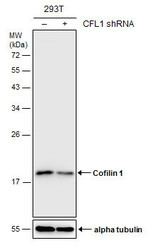
- Experimental details
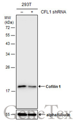
- Non-transfected (¡V) and transfected (+) 293T whole cell extracts (30 ?g) were separated by 12% SDS-PAGE, and the membrane was blotted with Cofilin 1 antibody (GTX102156) diluted at 1:1000. The HRP-conjugated anti-rabbit IgG antibody (GTX213110-01) was used to detect the primary antibody.
Supportive validation
- Submitted by
- GeneTex (provider)
- Main image
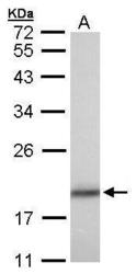
- Experimental details
- Sample (30 ?g of whole cell lysate) A: A431(GTX27909) 12% SDS PAGE GTX102156 diluted at 1:1000 The HRP-conjugated anti-rabbit IgG antibody (GTX213110-01) was used to detect the primary antibody.

- Submitted by
- GeneTex (provider)
- Main image

- Experimental details
- Sample (30 ?g of whole cell lysate) A:NIH-3T312% SDS PAGE GTX102156 diluted at 1:1000 The HRP-conjugated anti-rabbit IgG antibody (GTX213110-01) was used to detect the primary antibody.

- Submitted by
- GeneTex (provider)
- Main image

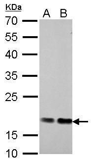
- Experimental details
- Cofilin 1 antibody detects CFL1 protein by western blot analysis.A. 30 ?g PC-12 whole cell lysate/extractB. 30 ?g Rat-2 whole cell lysate/extract12% SDS-PAGECofilin 1 antibody (GTX102156) dilution: 1:1000 The HRP-conjugated anti-rabbit IgG antibody (GTX213110-01) was used to detect the primary antibody.
- Submitted by
- GeneTex (provider)
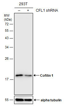
- Main image
- Experimental details
- Non-transfected (¡V) and transfected (+) 293T whole cell extracts (30 ?g) were separated by 12% SDS-PAGE, and the membrane was blotted with Cofilin 1 antibody (GTX102156) diluted at 1:1000. The HRP-conjugated anti-rabbit IgG antibody (GTX213110-01) was used to detect the primary antibody.

Supportive validation
- Submitted by
- GeneTex (provider)
- Main image
- Experimental details
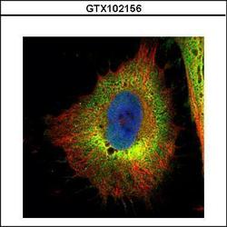
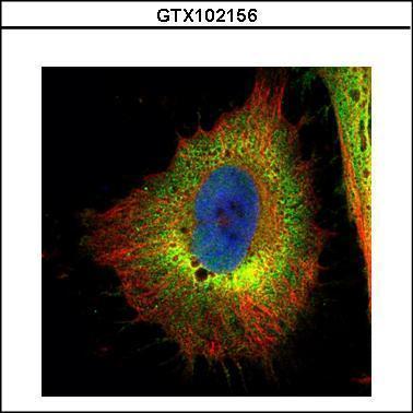
- Confocal immunofluorescence analysis (Olympus FV10i) of paraformaldehyde-fixed HeLa, using Cofilin 1 (non-muscle)(GTX102156) antibody (Green) at 1:500 dilution. Alpha-tubulin filaments were labeled with GTX11304 (Red) at 1:2000.
- Submitted by
- GeneTex (provider)
- Main image

- Experimental details
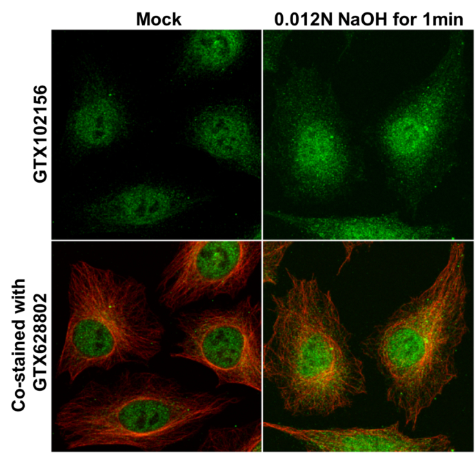
- Cofilin 1 antibody detects Cofilin 1 protein at cytoplasm and nucleus by immunofluorescent analysis. Samples: HeLa cells mock (left) and treated with 0.012 N NaOH/PBS for 1 min (right) were fixed in 4% paraformaldehyde at RT for 15 min.Green: Cofilin 1 protein stained by Cofilin 1 antibody (GTX102156) diluted at 1:1000.Red: alpha Tubulin, a cytoskeleton marker, stained by alpha Tubulin antibody [GT114] (GTX628802) diluted at 1:1000.
Supportive validation
- Submitted by
- GeneTex (provider)
- Main image
- Experimental details
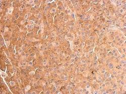
- Cofilin 1 antibody detects CFL1 protein at cytosol on HBL435 xenograft by immunohistochemical analysis. Sample: Paraffin-embedded HBL435 xenograft. Cofilin 1 antibody (GTX102156) dilution: 1:500.
