Explore
 Validate
 Learn
Learn Western blot
 Immunohistochemistry
Antibody data
- Antibody Data
- Antigen structure
- References [1]
- Comments [0]
- Validations
- Western blot [5]
- Immunocytochemistry [1]
- Immunohistochemistry [10]
Submit
Validation data
Reference
Comment
Report error
- Product number
- HPA020549 - Provider product page

- Provider
- Atlas Antibodies
- Proper citation
- Atlas Antibodies Cat#HPA020549, RRID:AB_1856834
- Product name
- Anti-SHMT2
- Antibody type
- Polyclonal
- Reactivity
- Human, Mouse, Rat
- Host
- Rabbit
- Conjugate
- Unconjugated
- Antigen sequence
NTCPGDRSAITPGGLRLGAPALTSRQFREDDFRRV
VDFIDEGVNIGLEVKSKTAKLQDFKSFLLKDSETS
QRLANLRQRVEQFARAF- Isotype
- IgG
- Vial size
- 100 µl
- Storage
- Store at +4°C for short term storage. Long time storage is recommended at -20°C.
Submitted references SHMT2 drives glioma cell survival in ischaemia but imposes a dependence on glycine clearance
Kim D, Fiske B, Birsoy K, Freinkman E, Kami K, Possemato R, Chudnovsky Y, Pacold M, Chen W, Cantor J, Shelton L, Gui D, Kwon M, Ramkissoon S, Ligon K, Kang S, Snuderl M, Vander Heiden M, Sabatini D
Nature 2015 April;520(7547):363-367
Nature 2015 April;520(7547):363-367
No comments: Submit comment
Supportive validation
Supportive validation
- Submitted by
- Atlas Antibodies (provider)
- Enhanced method
- Independent antibody validation
- Main image
- Experimental details
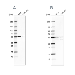
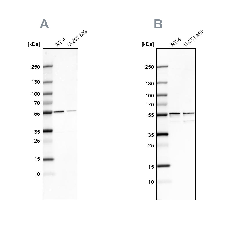
- Western blot analysis using Anti-SHMT2 antibody HPA020549 (A) shows similar pattern to independent antibody HPA020543 (B).
Supportive validation
- Submitted by
- Atlas Antibodies (provider)
- Main image
- Experimental details
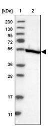
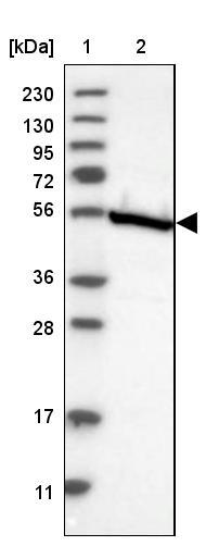
- Lane 1: Marker [kDa] 230, 130, 95, 72, 56, 36, 28, 17, 11Lane 2: Human cell line RT-4
- Sample type
- HUMAN
- Submitted by
- Atlas Antibodies (provider)
- Main image
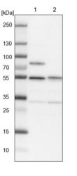
- Experimental details
- Lane 1: NIH-3T3 cell lysate (Mouse embryonic fibroblast cells)Lane 2: NBT-II cell lysate (Rat Wistar bladder tumour cells)

- Submitted by
- Atlas Antibodies (provider)
- Main image
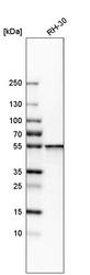
- Experimental details
- Western blot analysis in human cell line RH-30.

- Submitted by
- Atlas Antibodies (provider)
- Main image

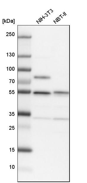
- Experimental details
- Western blot analysis in mouse cell line NIH-3T3 and rat cell line NBT-II.
Supportive validation
- Submitted by
- Atlas Antibodies (provider)
- Main image

- Experimental details
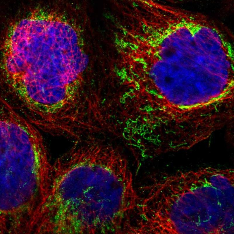
- Immunofluorescent staining of human cell line A-431 shows localization to mitochondria.
- Sample type
- HUMAN
Enhanced validation
Enhanced validation
Supportive validation
- Submitted by
- Atlas Antibodies (provider)
- Enhanced method
- Orthogonal validation
- Main image
- Experimental details
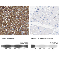
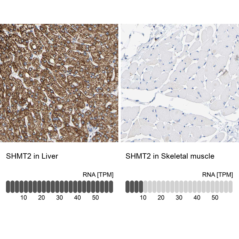
- Immunohistochemistry analysis in human liver and skeletal muscle tissues using HPA020549 antibody. Corresponding SHMT2 RNA-seq data are presented for the same tissues.
- Sample type
- HUMAN
Enhanced validation
- Submitted by
- Atlas Antibodies (provider)
- Enhanced method
- Independent antibody validation
- Main image
- Experimental details
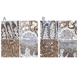
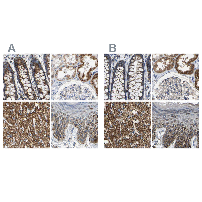
- Immunohistochemical staining of human colon, kidney, liver and skin using Anti-SHMT2 antibody HPA020549 (A) shows similar protein distribution across tissues to independent antibody HPA020543 (B).
Supportive validation
- Submitted by
- Atlas Antibodies (provider)
- Main image
- Experimental details
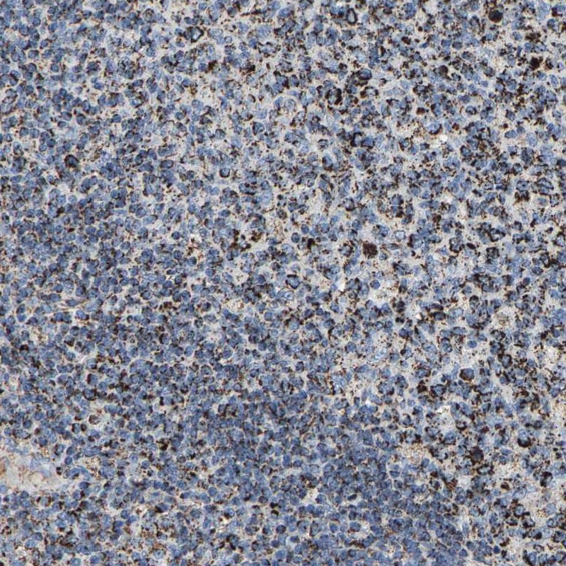
- Immunohistochemical staining of human lymph node shows strong cytoplasmic positivity in germinal center cells.
- Submitted by
- Atlas Antibodies (provider)
- Main image

- Experimental details
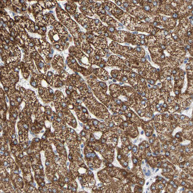
- Immunohistochemical staining of human liver shows moderate granular cytoplasmic positivity in hepatocytes.
- Sample type
- HUMAN
- Submitted by
- Atlas Antibodies (provider)
- Main image
- Experimental details
- Immunohistochemical staining of human lymph node shows moderate granular cytoplasmic positivity in germinal center cells.
- Sample type
- HUMAN

- Submitted by
- Atlas Antibodies (provider)
- Main image

- Experimental details
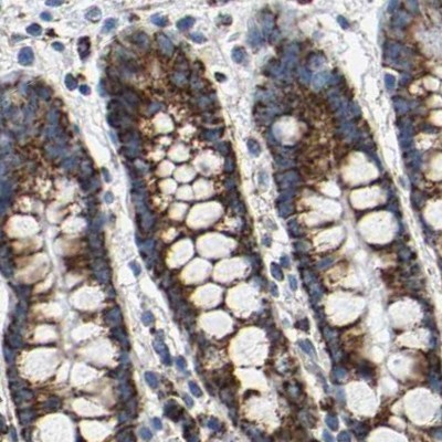
- Immunohistochemical staining of human stomach shows moderate granular cytoplasmic positivity in glandular cells.
- Sample type
- HUMAN
- Submitted by
- Atlas Antibodies (provider)
- Main image
- Experimental details
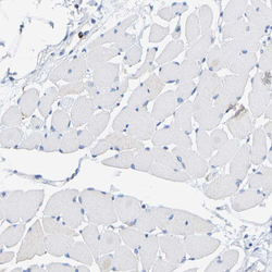
- Immunohistochemical staining of human skeletal muscle shows no positivity in myocytes as expected.
- Sample type
- HUMAN

- Submitted by
- Atlas Antibodies (provider)
- Main image

- Experimental details
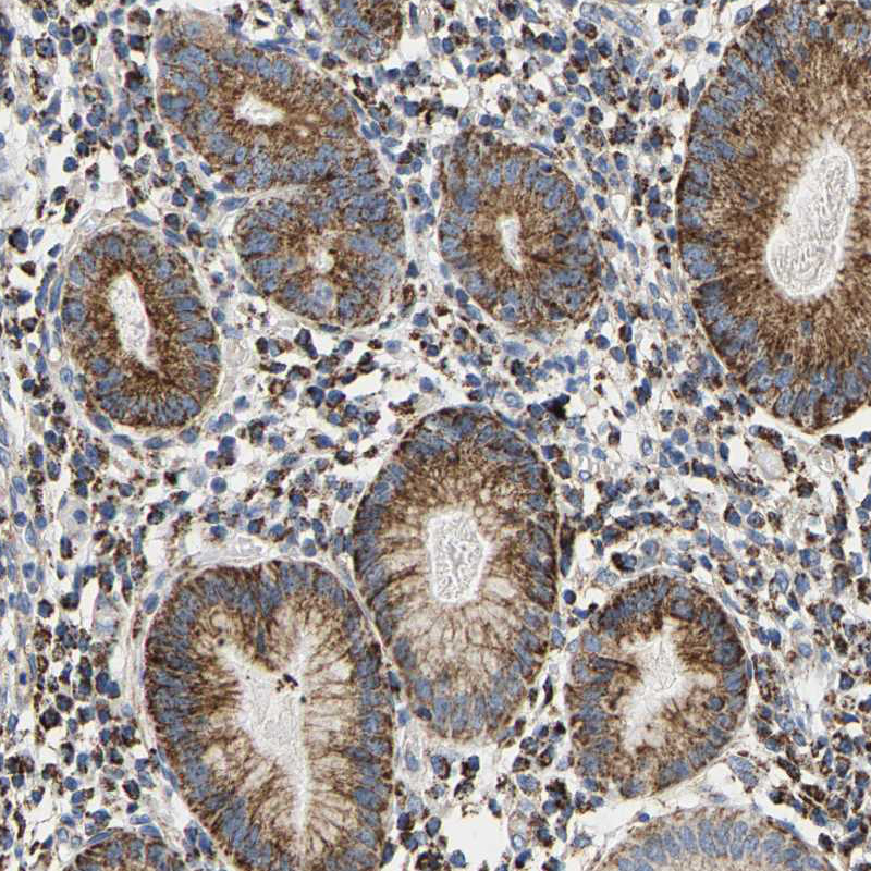
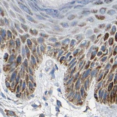
- Immunohistochemical staining of human colon using Anti-SHMT2 antibody HPA020549.
- Sample type
- HUMAN
- Submitted by
- Atlas Antibodies (provider)
- Main image

- Experimental details
- Immunohistochemical staining of human skin using Anti-SHMT2 antibody HPA020549.
- Sample type
- HUMAN
- Submitted by
- Atlas Antibodies (provider)
- Main image

- Experimental details
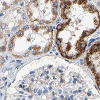
- Immunohistochemical staining of human kidney using Anti-SHMT2 antibody HPA020549.
- Sample type
- HUMAN