Explore
 Validate
 Learn
Learn Western blot
Antibody data
- Antibody Data
- Antigen structure
- References [0]
- Comments [0]
- Validations
- Western blot [2]
- Immunohistochemistry [1]
Submit
Validation data
Reference
Comment
Report error
- Product number
- APC-114-50UL - Provider product page

- Provider
- Invitrogen Antibodies
- Product name
- KCNH6 (erg2) Polyclonal Antibody
- Antibody type
- Polyclonal
- Antigen
- Other
- Reactivity
- Human, Mouse, Rat
- Host
- Rabbit
- Isotype
- IgG
- Vial size
- 50 µL
- Concentration
- 0.75 mg/mL
- Storage
- -20° C, Avoid Freeze/Thaw Cycles
No comments: Submit comment
Supportive validation
- Submitted by
- Invitrogen Antibodies (provider)
- Main image
- Experimental details
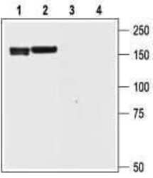
- Western blot analysisof rat brain (1 and 3) and cortex (2 and 4) lysate: - 1,2. Anti-KCNH6 (erg2) Antibody (#APC-114), (1:200).3,4. Anti-KCNH6 (erg2) Antibody , preincubated with KCNH6/erg2 Blocking Peptide (#BLP-PC114).

- Submitted by
- Invitrogen Antibodies (provider)
- Main image

- Experimental details
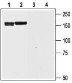
- Western blot analysisof rat brain (1 and 3) and cortex (2 and 4) lysate: - 1,2. Anti-KCNH6 (erg2) Antibody (#APC-114), (1:200).3,4. Anti-KCNH6 (erg2) Antibody , preincubated with KCNH6/erg2 Blocking Peptide (#BLP-PC114).
Supportive validation
- Submitted by
- Invitrogen Antibodies (provider)
- Main image
- Experimental details
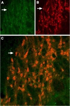
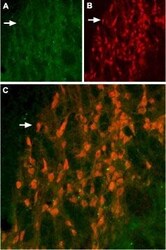
- Expression of KV11.2 (erg2) in rat hippocampus - Immunohistochemical staining of KV11.2 in rat hippocampus using Anti-KCNH6 (erg2) Antibody (#APC-114). A. KV11.2 appears as diffuse staining (green) that defines the boundary of the reticular nucleus of thalamus (arrow). B. Staining with mouse Anti-parvalbumin (PV, red) demonstrates both neuronal and diffuse staining. C. Confocal merge of KV11.2 and PV demonstrates overlap of the two markers labeling the reticular nucleus of the thalamus (orange).