 Explore
 Validate
 Learn
Learn Western blot
Antibody data
- Antibody Data
- Antigen structure
- References [1]
- Comments [0]
- Validations
- Western blot [2]
- Immunocytochemistry [1]
- Immunohistochemistry [3]
Submit
Validation data
Reference
Comment
Report error
- Product number
- MA5-37505 - Provider product page

- Provider
- Invitrogen Antibodies
- Product name
- TGF beta-2 Monoclonal Antibody (220ct16.4.3.1)
- Antibody type
- Monoclonal
- Antigen
- Recombinant full-length protein
- Reactivity
- Human
- Host
- Mouse
- Isotype
- IgG
- Antibody clone number
- 220ct16.4.3.1
- Vial size
- 400 µL
- Concentration
- 0.5 mg/mL
- Storage
- Store at 4°C short term. For long term storage, store at -20°C, avoiding freeze/thaw cycles.
Submitted references TGF-β1 mediates pathologic changes of secondary lymphedema by promoting fibrosis and inflammation.
Baik JE, Park HJ, Kataru RP, Savetsky IL, Ly CL, Shin J, Encarnacion EM, Cavali MR, Klang MG, Riedel E, Coriddi M, Dayan JH, Mehrara BJ
Clinical and translational medicine 2022 Jun;12(6):e758
Clinical and translational medicine 2022 Jun;12(6):e758
No comments: Submit comment
Supportive validation
- Submitted by
- Invitrogen Antibodies (provider)
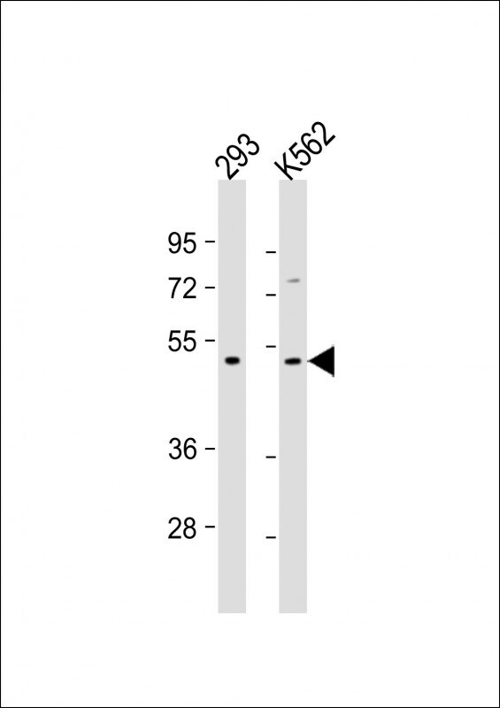
- Main image

- Experimental details
- Western Blot analysis of TGF beta-2 using the TGF beta-2 Monoclonal Antibody (Product # MA5-37505) at a 1:500-1:1000 dilution. Lane 1: 293 whole cell lysate, Lane 2: K562 whole cell lysate. Lysates/proteins were loaded at 20 µg per lane. Secondary Goat Anti-mouse IgG, (H+L), Peroxidase conjugated at 1:10,000 dilution. Predicted band size : 48 kDa. Blocking/Dilution buffer: 5% NFDM/TBST.
- Submitted by
- Invitrogen Antibodies (provider)
- Main image

- Experimental details
- Western Blot analysis of TGF beta-2 using the TGF beta-2 Monoclonal Antibody (Product # MA5-37505) in A549 cell line lysates (35 µg/lane).

Supportive validation
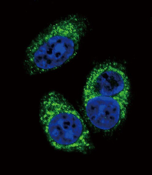
- Submitted by
- Invitrogen Antibodies (provider)
- Main image

- Experimental details
- Immunocytochemistry/Immunofluorescent analysis of TGF beta-2 in A549 cells using the TGF beta-2 Monoclonal Antibody (Product # MA5-37505) at a dilution of 1:10-1:50, followed by Alexa Fluor® 488-conjugated goat anti-mouse lgG (green).DAPI was used to stain the cell nuclear (blue).
Supportive validation
- Submitted by
- Invitrogen Antibodies (provider)
- Main image

- Experimental details
- Immunohistochemistry (Paraffin) analysis of TGF beta-2 in paraffin-embedded Human kidney sections using the TGF beta-2 Monoclonal Antibody (Product # MA5-37505) at a dilution of 1:50. A undiluted biotinylated goat polyvalent antibody was used as the secondary, followed by DAB staining.

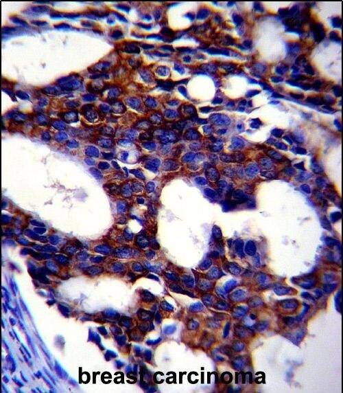
- Submitted by
- Invitrogen Antibodies (provider)
- Main image

- Experimental details
- Immunohistochemistry (Paraffin) analysis of TGF beta-2 in formalin fixed and paraffin embedded human breast carcinoma tissue using the TGF beta-2 Monoclonal Antibody (Product # MA5-37505) at a dilution of 1:10-1:50, followed by peroxidase conjugation of the secondary antibody and DAB staining.
- Submitted by
- Invitrogen Antibodies (provider)
- Main image
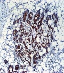
- Experimental details
- Immunohistochemistry (Paraffin) analysis of TGF beta-2 in formalin fixed and paraffin embedded human colon carcinoma tissue using the TGF beta-2 Monoclonal Antibody (Product # MA5-37505) at a dilution of 1:10-1:50, followed by peroxidase conjugation of the secondary antibody and DAB staining.
