 Explore
 Validate
 Learn
Learn Western blot
Antibody data
- Antibody Data
- Antigen structure
- References [5]
- Comments [0]
- Validations
- Western blot [1]
- Immunohistochemistry [1]
Submit
Validation data
Reference
Comment
Report error
- Product number
- AF5806 - Provider product page

- Provider
- Novus Biologicals
- Product name
- Goat Polyclonal Siglec-E Antibody
- Antibody type
- Polyclonal
- Description
- Antigen Affinity-purified. Detects mouse Siglec-E in direct ELISAs and Western blots. In direct ELISAs, less than 3% cross-reactivity with recombinant human (rh) Siglec-6, rhSiglec-7, and rhSiglec-9 is observed.
- Reactivity
- Mouse
- Host
- Goat
- Conjugate
- Unconjugated
- Isotype
- IgG
- Vial size
- 100 ug
- Concentration
- LYOPH
- Storage
- Use a manual defrost freezer and avoid repeated freeze-thaw cycles. 12 months from date of receipt, -20 to -70 degreesC as supplied. 1 month, 2 to 8 degreesC under sterile conditions after reconstitution. 6 months, -20 to -70 degreesC under sterile conditions after reconstitution.
Submitted references Studies on the Detection, Expression, Glycosylation, Dimerization, and Ligand Binding Properties of Mouse Siglec-E.
Siglec-7 restores β-cell function and survival and reduces inflammation in pancreatic islets from patients with diabetes.
Leishmania donovani Utilize Sialic Acids for Binding and Phagocytosis in the Macrophages through Selective Utilization of Siglecs and Impair the Innate Immune Arm.
Siglec receptors impact mammalian lifespan by modulating oxidative stress.
Group B Streptococcus engages an inhibitory Siglec through sialic acid mimicry to blunt innate immune and inflammatory responses in vivo.
Siddiqui S, Schwarz F, Springer S, Khedri Z, Yu H, Deng L, Verhagen A, Naito-Matsui Y, Jiang W, Kim D, Zhou J, Ding B, Chen X, Varki N, Varki A
The Journal of biological chemistry 2017 Jan 20;292(3):1029-1037
The Journal of biological chemistry 2017 Jan 20;292(3):1029-1037
Siglec-7 restores β-cell function and survival and reduces inflammation in pancreatic islets from patients with diabetes.
Dharmadhikari G, Stolz K, Hauke M, Morgan NG, Varki A, de Koning E, Kelm S, Maedler K
Scientific reports 2017 Apr 5;7:45319
Scientific reports 2017 Apr 5;7:45319
Leishmania donovani Utilize Sialic Acids for Binding and Phagocytosis in the Macrophages through Selective Utilization of Siglecs and Impair the Innate Immune Arm.
Roy S, Mandal C
PLoS neglected tropical diseases 2016 Aug;10(8):e0004904
PLoS neglected tropical diseases 2016 Aug;10(8):e0004904
Siglec receptors impact mammalian lifespan by modulating oxidative stress.
Schwarz F, Pearce OM, Wang X, Samraj AN, Läubli H, Garcia JO, Lin H, Fu X, Garcia-Bingman A, Secrest P, Romanoski CE, Heyser C, Glass CK, Hazen SL, Varki N, Varki A, Gagneux P
eLife 2015 Apr 7;4
eLife 2015 Apr 7;4
Group B Streptococcus engages an inhibitory Siglec through sialic acid mimicry to blunt innate immune and inflammatory responses in vivo.
Chang YC, Olson J, Beasley FC, Tung C, Zhang J, Crocker PR, Varki A, Nizet V
PLoS pathogens 2014 Jan;10(1):e1003846
PLoS pathogens 2014 Jan;10(1):e1003846
No comments: Submit comment
Supportive validation
- Submitted by
- Novus Biologicals (provider)
- Main image
- Experimental details
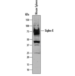
- Detection of Mouse Siglec-E by Western Blot. Western blot shows lysates of mouse spleen tissue. PVDF membrane was probed with 1 µg/mL of Goat Anti-Mouse Siglec-E Antigen Affinity-purified Polyclonal Antibody (Catalog # AF5806) followed by HRP-conjugated Anti-Goat IgG Secondary Antibody (Catalog # HAF109). A specific band was detected for Siglec-E at approximately 80 kDa (as indicated). This experiment was conducted under reducing conditions and using Immunoblot Buffer Group 1.

Supportive validation
- Submitted by
- Novus Biologicals (provider)
- Main image
- Experimental details
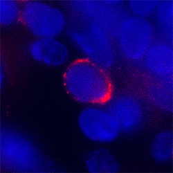
- Siglec-E in Mouse Spleen. Siglec-E was detected in perfusion fixed frozen sections of mouse spleen using Goat Anti-Mouse Siglec-E Antigen Affinity-purified Polyclonal Antibody (Catalog # AF5806) at 1.7 µg/mL overnight at 4 °C. Tissue was stained using the NorthernLights™ 557-conjugated Anti-Goat IgG Secondary Antibody (red; Catalog # NL001) and counterstained with DAPI (blue). Specific staining was localized to plasma membrane. View our protocol for Fluorescent IHC Staining of Frozen Tissue Sections.
