Antibody data
- Antibody Data
- Antigen structure
- References [1]
- Comments [0]
- Validations
- Western blot [4]
- Immunohistochemistry [1]
- Other assay [1]
Submit
Validation data
Reference
Comment
Report error
- Product number
- PA5-79118 - Provider product page

- Provider
- Invitrogen Antibodies
- Product name
- NOX2 Polyclonal Antibody
- Antibody type
- Polyclonal
- Antigen
- Synthetic peptide
- Description
- Reconstitute with 0.2 mL of distilled water to yield a concentration of 500 µg/mL.
- Reactivity
- Human, Mouse, Rat
- Host
- Rabbit
- Isotype
- IgG
- Vial size
- 100 µg
- Concentration
- 500 µg/mL
- Storage
- -20°C
Submitted references Activated Histone Acetyltransferase p300/CBP-Related Signalling Pathways Mediate Up-Regulation of NADPH Oxidase, Inflammation, and Fibrosis in Diabetic Kidney.
Lazar AG, Vlad ML, Manea A, Simionescu M, Manea SA
Antioxidants (Basel, Switzerland) 2021 Aug 26;10(9)
Antioxidants (Basel, Switzerland) 2021 Aug 26;10(9)
No comments: Submit comment
Supportive validation
- Submitted by
- Invitrogen Antibodies (provider)
- Main image
- Experimental details
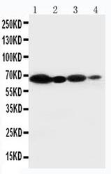
- Western blot analysis of NOX2 in Lane 1: HeLa cell lysate, Lane 2: JURKAT cell lysate, Lane 3: MCF-7 cell lysate, Lane 4: SMMC cell lysate. Sample was incubated with NOX2 polyclonal antibody (Product # PA5-79118).
- Submitted by
- Invitrogen Antibodies (provider)
- Main image
- Experimental details
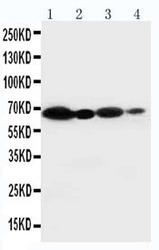
- Western blot analysis of NOX2 in Lane 1: HeLa cell lysate, Lane 2: JURKAT cell lysate, Lane 3: MCF-7 cell lysate, Lane 4: SMMC cell lysate. Sample was incubated with NOX2 polyclonal antibody (Product # PA5-79118).
- Submitted by
- Invitrogen Antibodies (provider)
- Main image
- Experimental details
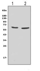
- Western blot analysis of NOX2 in, Lane 1: rat thymus tissue lysates, Lane 2: rat brain tissue lysates, . Electrophoresis was performed on a 5-20% SDS-PAGE gel at 70V (Stacking gel) / 90V (Resolving gel) for 2-3 hours. The sample well of each lane was loaded with 50 µg of sample under reducing conditions. After Electrophoresis, proteins were transferred to a Nitrocellulose membrane at 150mA for 50-90 minutes. The membrane was blocked with 5% Non-fat Milk/ TBS for 1.5 hour at RT. The membrane was incubated with NOX2 Polyclonal Antibody (Product # PA5-79118) at 0.5 μg/mL overnight at 4°C, then washed with TBS-0.1%Tween 3 times with 5 minutes each and probed with a goat anti-rabbit IgG-HRP secondary antibody at a dilution of 1:10000 for 1.5 hour at RT. The signal is developed using an Enhanced Chemiluminescent detection (ECL) kit. A specific band was detected for NOX2 at approximately 65KD. The expected band size for NOX2 is at 65KD.
- Submitted by
- Invitrogen Antibodies (provider)
- Main image

- Experimental details
- Western blot was performed using Anti-NOX2 Polyclonal Antibody (Product # PA5-79118) and a 58 kDa, 63 kDa band corresponding to NOX2 was observed across cell lines. Tissue extraction buffer extracts (30 µg lysate) of Raji (Lane 1), Daudi (Lane 2), Jurkat (Lane 3), SK-O-V3 (Lane 4) and HeLa (Lane 5) were electrophoresed using NuPAGE™ 4-12% Bis-Tris Protein Gel (Product # NP0322BOX). Resolved proteins were then transferred onto a nitrocellulose membrane (Product # IB23001) by iBlot® 2 Dry Blotting System (Product # IB21001). The blot was probed with the primary antibody (1:1000 dilution) and detected by chemiluminescence with Goat anti-Rabbit IgG (H+L), Superclonal™ Recombinant Secondary Antibody, HRP (Product # A27036, 1:4000 dilution) using the iBright FL 1000 (Product # A32752). Chemiluminescent detection was performed using Novex® ECL Chemiluminescent Substrate Reagent Kit (Product # WP20005).
Supportive validation
- Submitted by
- Invitrogen Antibodies (provider)
- Main image
- Experimental details
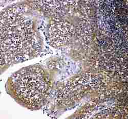
- Immunohistochemistry analysis of NOX2 on paraffin-embedded human intestinal cancer tissue. Sample was incubated with NOX2 polyclonal antibody (Product# PA5-79118).
Supportive validation
- Submitted by
- Invitrogen Antibodies (provider)
- Main image
- Experimental details
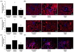
- Figure 4 Pharmacological inhibition of histone acetyltransferase p300/CBP by C646 mitigates the glomerular expression of Nox subtypes in diabetic mice. ( A , C , E ) Quantification of fluorescence immunolabeling for glomerular Nox1, Nox2, and Nox4 subtypes. ( B , D , F ) Representative IF microscopy images taken at 40x magnification depicting the immunofluorescence staining (red) for Nox subtypes. Sections were counterstained with DAPI (blue) stain to detect the cell nuclei in the specimens. Note that the up-regulation of glomerular Nox1, Nox2, and Nox4 protein levels in diabetic mice is blunted following C646 treatment. n = 14-19/condition quantified glomeruli, *** p < 0.001, p -values taken in relation to non-diabetic + vehicle condition; ### p < 0.001, p -values taken in relation to diabetic + vehicle condition.
 Explore
 Validate
 Learn
Learn Western blot