 Explore
 Validate
 Learn
Learn Western blot
Antibody data
- Antibody Data
- Antigen structure
- References [22]
- Comments [0]
- Validations
- Western blot [1]
- Immunocytochemistry [1]
- Flow cytometry [1]
- Other assay [14]
Submit
Validation data
Reference
Comment
Report error
- Product number
- PA1-660 - Provider product page

- Provider
- Invitrogen Antibodies
- Product name
- Dynamin 1 Polyclonal Antibody
- Antibody type
- Polyclonal
- Antigen
- Synthetic peptide
- Description
- PA1-660 detects dynamin I (Dyn1) from rat tissues. PA1-660 has been successfully used in Western blot and immunocytochemical procedures. By Western blot, this antibody detects an ~100 kDa protein representing Dyn1 from rat whole brain extract. PA1-660 immunizing peptide corresponds to amino acid residues 633-647 from rat Dyn1. This sequence is completely conserved between rat, mouse, and human Dyn1. PA1-660 immunizing peptide (Cat. # PEP-042) is available for use in neutralization and control experiments.
- Reactivity
- Human, Mouse, Rat
- Host
- Rabbit
- Isotype
- IgG
- Vial size
- 100 µL
- Concentration
- 1 mg/mL
- Storage
- -20° C, Avoid Freeze/Thaw Cycles
Submitted references Dopaminergic neurodegeneration induced by Parkinson's disease-linked G2019S LRRK2 is dependent on kinase and GTPase activity.
The invasive proteome of glioblastoma revealed by laser-capture microdissection.
Dynamin 1 Restrains Vesicular Release to a Subquantal Mode In Mammalian Adrenal Chromaffin Cells.
JC Polyomavirus Entry by Clathrin-Mediated Endocytosis Is Driven by β-Arrestin.
PICK1 regulates AMPA receptor endocytosis via direct interactions with AP2 α-appendage and dynamin.
Actin bundling by dynamin 2 and cortactin is implicated in cell migration by stabilizing filopodia in human non-small cell lung carcinoma cells.
Dynamin 1 isoform roles in a mouse model of severe childhood epileptic encephalopathy.
Independent Neuronal Origin of Seizures and Behavioral Comorbidities in an Animal Model of a Severe Childhood Genetic Epileptic Encephalopathy.
Dynamin 1 is required for memory formation.
Synaptic function is modulated by LRRK2 and glutamate release is increased in cortical neurons of G2019S LRRK2 knock-in mice.
Parkinson's disease-linked mutations in VPS35 induce dopaminergic neurodegeneration.
Alzheimer brain-derived amyloid β-protein impairs synaptic remodeling and memory consolidation.
Stabilization of actin bundles by a dynamin 1/cortactin ring complex is necessary for growth cone filopodia.
Protein partners of dynamin-1 in the retina.
Endocytosis in cultured neurons is altered by chronic alcohol exposure.
Dynamin 1 depletion and memory deficits in rats treated with Abeta and cerebral ischemia.
A missense mutation in a highly conserved alternate exon of dynamin-1 causes epilepsy in fitful mice.
Overlapping functions of different dynamin isoforms in clathrin-dependent and -independent endocytosis in pancreatic beta cells.
A selective activity-dependent requirement for dynamin 1 in synaptic vesicle endocytosis.
Beta-amyloid-induced dynamin 1 depletion in hippocampal neurons. A potential mechanism for early cognitive decline in Alzheimer disease.
Phosphatidylinositol 4,5-bisphosphate stimulates vesicle formation from liposomes by brain cytosol.
Aquaporin-2 localization in clathrin-coated pits: inhibition of endocytosis by dominant-negative dynamin.
Nguyen APT, Tsika E, Kelly K, Levine N, Chen X, West AB, Boularand S, Barneoud P, Moore DJ
Proceedings of the National Academy of Sciences of the United States of America 2020 Jul 21;117(29):17296-17307
Proceedings of the National Academy of Sciences of the United States of America 2020 Jul 21;117(29):17296-17307
The invasive proteome of glioblastoma revealed by laser-capture microdissection.
Daubon T, Guyon J, Raymond AA, Dartigues B, Rudewicz J, Ezzoukhry Z, Dupuy JW, Herbert JMJ, Saltel F, Bjerkvig R, Nikolski M, Bikfalvi A
Neuro-oncology advances 2019 May-Dec;1(1):vdz029
Neuro-oncology advances 2019 May-Dec;1(1):vdz029
Dynamin 1 Restrains Vesicular Release to a Subquantal Mode In Mammalian Adrenal Chromaffin Cells.
Wu Q, Zhang Q, Liu B, Li Y, Wu X, Kuo S, Zheng L, Wang C, Zhu F, Zhou Z
The Journal of neuroscience : the official journal of the Society for Neuroscience 2019 Jan 9;39(2):199-211
The Journal of neuroscience : the official journal of the Society for Neuroscience 2019 Jan 9;39(2):199-211
JC Polyomavirus Entry by Clathrin-Mediated Endocytosis Is Driven by β-Arrestin.
Mayberry CL, Soucy AN, Lajoie CR, DuShane JK, Maginnis MS
Journal of virology 2019 Apr 15;93(8)
Journal of virology 2019 Apr 15;93(8)
PICK1 regulates AMPA receptor endocytosis via direct interactions with AP2 α-appendage and dynamin.
Fiuza M, Rostosky CM, Parkinson GT, Bygrave AM, Halemani N, Baptista M, Milosevic I, Hanley JG
The Journal of cell biology 2017 Oct 2;216(10):3323-3338
The Journal of cell biology 2017 Oct 2;216(10):3323-3338
Actin bundling by dynamin 2 and cortactin is implicated in cell migration by stabilizing filopodia in human non-small cell lung carcinoma cells.
Yamada H, Takeda T, Michiue H, Abe T, Takei K
International journal of oncology 2016 Sep;49(3):877-86
International journal of oncology 2016 Sep;49(3):877-86
Dynamin 1 isoform roles in a mouse model of severe childhood epileptic encephalopathy.
Asinof S, Mahaffey C, Beyer B, Frankel WN, Boumil R
Neurobiology of disease 2016 Nov;95:1-11
Neurobiology of disease 2016 Nov;95:1-11
Independent Neuronal Origin of Seizures and Behavioral Comorbidities in an Animal Model of a Severe Childhood Genetic Epileptic Encephalopathy.
Asinof SK, Sukoff Rizzo SJ, Buckley AR, Beyer BJ, Letts VA, Frankel WN, Boumil RM
PLoS genetics 2015 Jun;11(6):e1005347
PLoS genetics 2015 Jun;11(6):e1005347
Dynamin 1 is required for memory formation.
Fà M, Staniszewski A, Saeed F, Francis YI, Arancio O
PloS one 2014;9(3):e91954
PloS one 2014;9(3):e91954
Synaptic function is modulated by LRRK2 and glutamate release is increased in cortical neurons of G2019S LRRK2 knock-in mice.
Beccano-Kelly DA, Kuhlmann N, Tatarnikov I, Volta M, Munsie LN, Chou P, Cao LP, Han H, Tapia L, Farrer MJ, Milnerwood AJ
Frontiers in cellular neuroscience 2014;8:301
Frontiers in cellular neuroscience 2014;8:301
Parkinson's disease-linked mutations in VPS35 induce dopaminergic neurodegeneration.
Tsika E, Glauser L, Moser R, Fiser A, Daniel G, Sheerin UM, Lees A, Troncoso JC, Lewis PA, Bandopadhyay R, Schneider BL, Moore DJ
Human molecular genetics 2014 Sep 1;23(17):4621-38
Human molecular genetics 2014 Sep 1;23(17):4621-38
Alzheimer brain-derived amyloid β-protein impairs synaptic remodeling and memory consolidation.
Borlikova GG, Trejo M, Mably AJ, Mc Donald JM, Sala Frigerio C, Regan CM, Murphy KJ, Masliah E, Walsh DM
Neurobiology of aging 2013 May;34(5):1315-27
Neurobiology of aging 2013 May;34(5):1315-27
Stabilization of actin bundles by a dynamin 1/cortactin ring complex is necessary for growth cone filopodia.
Yamada H, Abe T, Satoh A, Okazaki N, Tago S, Kobayashi K, Yoshida Y, Oda Y, Watanabe M, Tomizawa K, Matsui H, Takei K
The Journal of neuroscience : the official journal of the Society for Neuroscience 2013 Mar 6;33(10):4514-26
The Journal of neuroscience : the official journal of the Society for Neuroscience 2013 Mar 6;33(10):4514-26
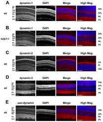
Protein partners of dynamin-1 in the retina.
Grossman GH, Ebke LA, Beight CD, Jang GF, Crabb JW, Hagstrom SA
Visual neuroscience 2013 Jul;30(4):129-39
Visual neuroscience 2013 Jul;30(4):129-39
Endocytosis in cultured neurons is altered by chronic alcohol exposure.
Marín MP, Esteban-Pretel G, Ponsoda X, Romero AM, Ballestín R, López C, Megías L, Timoneda J, Molowny A, Canales JJ, Renau-Piqueras J
Toxicological sciences : an official journal of the Society of Toxicology 2010 May;115(1):202-13
Toxicological sciences : an official journal of the Society of Toxicology 2010 May;115(1):202-13
Dynamin 1 depletion and memory deficits in rats treated with Abeta and cerebral ischemia.
Watanabe T, Iwasaki K, Takasaki K, Yamagata N, Fujino M, Nogami A, Ii M, Katsurabayashi S, Mishima K, Fujiwara M
Journal of neuroscience research 2010 Jul;88(9):1908-17
Journal of neuroscience research 2010 Jul;88(9):1908-17
A missense mutation in a highly conserved alternate exon of dynamin-1 causes epilepsy in fitful mice.
Boumil RM, Letts VA, Roberts MC, Lenz C, Mahaffey CL, Zhang ZW, Moser T, Frankel WN
PLoS genetics 2010 Aug 5;6(8)
PLoS genetics 2010 Aug 5;6(8)
Overlapping functions of different dynamin isoforms in clathrin-dependent and -independent endocytosis in pancreatic beta cells.
Lu J, He Z, Fan J, Xu P, Chen L
Biochemical and biophysical research communications 2008 Jun 27;371(2):315-9
Biochemical and biophysical research communications 2008 Jun 27;371(2):315-9
A selective activity-dependent requirement for dynamin 1 in synaptic vesicle endocytosis.
Ferguson SM, Brasnjo G, Hayashi M, Wölfel M, Collesi C, Giovedi S, Raimondi A, Gong LW, Ariel P, Paradise S, O'toole E, Flavell R, Cremona O, Miesenböck G, Ryan TA, De Camilli P
Science (New York, N.Y.) 2007 Apr 27;316(5824):570-4
Science (New York, N.Y.) 2007 Apr 27;316(5824):570-4
Beta-amyloid-induced dynamin 1 depletion in hippocampal neurons. A potential mechanism for early cognitive decline in Alzheimer disease.
Kelly BL, Vassar R, Ferreira A
The Journal of biological chemistry 2005 Sep 9;280(36):31746-53
The Journal of biological chemistry 2005 Sep 9;280(36):31746-53
Phosphatidylinositol 4,5-bisphosphate stimulates vesicle formation from liposomes by brain cytosol.
Kinuta M, Yamada H, Abe T, Watanabe M, Li SA, Kamitani A, Yasuda T, Matsukawa T, Kumon H, Takei K
Proceedings of the National Academy of Sciences of the United States of America 2002 Mar 5;99(5):2842-7
Proceedings of the National Academy of Sciences of the United States of America 2002 Mar 5;99(5):2842-7
Aquaporin-2 localization in clathrin-coated pits: inhibition of endocytosis by dominant-negative dynamin.
Sun TX, Van Hoek A, Huang Y, Bouley R, McLaughlin M, Brown D
American journal of physiology. Renal physiology 2002 Jun;282(6):F998-1011
American journal of physiology. Renal physiology 2002 Jun;282(6):F998-1011
No comments: Submit comment
Supportive validation
- Submitted by
- Invitrogen Antibodies (provider)
- Main image

- Experimental details
- Western blot analysis was performed using whole cell extract (30 µg) of RAW 264.7 (Lane 1), PC-12 (Lane 2), PC-12 treated with Retinoic acid 1uM for 3 days (Lane 3), SH-SY5Y (Lane 4) and SH-SY5Y treated with Retinoic acid 1uM for 3 days (Lane 5). The blots were probed with Anti- Dynamin I Rabbit Polyclonal (Product # PA1-660, 2 µg/mL) and detected by chemiluminescence using Goat anti-Rabbit IgG (H+L) Superclonal™ Secondary Antibody, HRP conjugate (Product # A27036, 0.4 µg/mL, 1:2500 dilution). A ~ 97 kDa band corresponding to Dynamin I was observed across cell lines tested. Known quantity of protein samples were electrophoresed using Novex® NuPAGE® 4-12 % Bis-Tris gel (Product # NP0321BOX), XCell SureLock™ Electrophoresis System (Product # EI0002) and Novex® Sharp Pre-Stained Protein Standard (Product # LC5800).) Resolved proteins were then transferred onto a nitrocellulose membrane by Pierce™ Power Blotter System (22834). The membrane was probed with the relevant primary and secondary Antibody following blocking with 5 % skimmed milk. Chemiluminescent detection was performed using Pierce™ ECL Western Blotting Substrate (Product # 32106).

Supportive validation
- Submitted by
- Invitrogen Antibodies (provider)
- Main image

- Experimental details
- Immunofluorescence analysis of Dynamin I was performed using 70% confluent log phase SH-SY5Y cells. The cells were fixed with 4% paraformaldehyde for 10 minutes, permeabilized with 0.1% Triton™ X-100 for 10 minutes, and blocked with 1% BSA for 1 hour at room temperature. The cells were labeled with Dynamin I Rabbit Polyclonal Antibody (Product # PA1-660) at 2µg/mL in 0.1% BSA and incubated for 3 hours at room temperature and then labeled with Goat anti-Rabbit IgG (H+L) Superclonal™ Secondary Antibody, Alexa Fluor® 488 conjugate (Product # A27034) a dilution of 1:2000 for 45 minutes at room temperature (Panel a: green). Nuclei (Panel b: blue) were stained with SlowFade® Gold Antifade Mountant with DAPI (Product # S36938). F-actin (Panel c: red) was stained with Alexa Fluor® 555 Rhodamine Phalloidin (Product # R415, 1:300). Panel d represents the merged image showing cytoplasmic localization. Panel e shows the no primary antibody control. The images were captured at 60X magnification.

Supportive validation
- Submitted by
- Invitrogen Antibodies (provider)
- Main image

- Experimental details
- Flow cytometry analysis of Dynamin I was performed using SH-SY5Y cells. Cells were fixed with 70% ethanol for 10 minutes, permeabilized with 0.25% Triton™ X-100 for 20 minutes, and blocked with 5% BSA for 30 minutes at room temperature. Cells were labeled with Dynamin I Rabbit Polyclonal Antibody (Product # PA1-660, red histogram) or with rabbit isotype control (pink histogram) at 3-5 µg/million cells in 2.5% BSA. After incubation at room temperature for 2 hours, the cells were labeled with Alexa Fluor® 488 Goat Anti-Rabbit Secondary Antibody (Product # A11008) at a dilution of 1:400 for 30 minutes at room temperature. The representative 10, 000 cells were acquired and analyzed for each sample using an Attune® Acoustic Focusing Cytometer. The purple histogram represents unstained control cells and the green histogram represents no-primary-antibody control.

Supportive validation
- Submitted by
- Invitrogen Antibodies (provider)
- Main image

- Experimental details
- NULL

- Submitted by
- Invitrogen Antibodies (provider)
- Main image

- Experimental details
- NULL

- Submitted by
- Invitrogen Antibodies (provider)
- Main image

- Experimental details
- NULL

- Submitted by
- Invitrogen Antibodies (provider)
- Main image
- Experimental details
- NULL

- Submitted by
- Invitrogen Antibodies (provider)
- Main image

- Experimental details
- NULL

- Submitted by
- Invitrogen Antibodies (provider)
- Main image

- Experimental details
- NULL

- Submitted by
- Invitrogen Antibodies (provider)
- Main image

- Experimental details
- NULL

- Submitted by
- Invitrogen Antibodies (provider)
- Main image

- Experimental details
- Figure 5 DNM1a Ftfl is defective in higher order homo-oligomerization. (A) Left panel , protein extract from P14 whole brain tissue of homozygous fitful and wildtype littermates incubated with 0 or 20mM EDC cross-linker and hybridized with anti-dynamin-1 antibody. Monomers migrate at 100kD, dimers at 200kD and the tetramers are at 400kD. This assay was performed over three separate times with different samples each time; a representative blot with corresponding percentages is shown. Mean densities (+- 1SD) from all experiments are: wildtype 28.75+-8.24 (monomer), 29.67+-13.9 (dimer), 43.9+-8.5 (tetramer); mutant 44+-10.7 (monomer), 39.5+-12.2 (dimer), 23+-13.5 (tetramer) Right panel , COS-7 cells transfected with DNM1-GFP constructs show differences in dimerization. (B) COS-7 cells doubly transfected with DNM1-GFP and DNM1-HA constructs show isoform heterodimerization. Protein extracts from cells were incubated with 0 or 20mM EDC and analyzed by Western blot. Blots were hybridized with anti-GFP antibody, stripped of antibody and then re-hybridized with anti-HA antibody in order to ascertain the presence of each construct in the dimers. A representative blot hybridized with anti-GFP antibody is shown.

- Submitted by
- Invitrogen Antibodies (provider)
- Main image

- Experimental details
- Figure 4 Reduced Synapsin 1 phosphorylation in KI cortical neurons . Levels of pre-synaptic proteins in DIV21 CTX cultures were assayed by standard western blotting and verified via WES automated capillary-based size sorting system. (A) Representative western blots of EndophilinA (EndoA), vesicle associated membrane protein 1 (VAMP1), vesicle associated membrane protein 2 (VAMP2), dynamin 1, synapsin1 (Syn1), phosphoserine 9 synapsin1 (pS9 Syn1), and phosphoserine 603 synapsin1 (pS603 Syn1). (B) Quantification of synapsin1 levels and associated phosphorylation sites. Synapsin1 levels were similar between NT and KI however the ratio of phosphorylated synapsin1 was significantly reduced at both sites. (C) Standard western blot results were verified using the WES automated capillary-based size sorting system for the S603 phosphorylation site. Representative pseudo-gels (left) and electropherograms (right) exported from the WES compass analysis software. (D) Quantification of synapsin1 and pS603 synapsin1 confirmed significant reductions pS603 synapsin1. Data expressed relative to GAPDH and normalized to NT, * p

- Submitted by
- Invitrogen Antibodies (provider)
- Main image

- Experimental details
- Figure 6 Selective dynamin 1 inhibition through siRNA impairs both synaptic plasticity and associative memory. A , siRNA specific for murine dynamin 1 reduces protein expression. An example of western blot showing that Penetratin 1- conjugated dynamin 1 siRNA reduces protein expression. Cells are lysed 48 hours after the treatment with siRNA. Dynamin 1 is detected using a rabbit polyclonal anti-dynamin1 antibody. Penetratin 1- conjugated Control siRNA, that does not affect dynamin 1 expression, does not change protein levels. n = 3 for each group. B , Penetratin 1- conjugated dynamin 1 siRNA (open bars) (80 nM in a final volume of 1.5 mul over 1 minute, bilateral injections twice a day for 3 days) impairs contextual fear memory compared to control siRNA infused mice (grey bars) (Mann-Whitney U 150, 126 = 21.00, p = 0.0089). Moreover, control siRNA infused mice show similar amount of freezing as vehicle-infused animals (black bars). C , Penetratin 1- conjugated dynamin 1 siRNA (open bars) does not modify cued fear memory compared to Penetratin 1- conjugated Control siRNA (grey bars) (Mann-Whitney U 99, 90 = 44.50, p = 1.00) in mice previously tested for contextual fear conditioning at 24 hours after the shock. D , Bilateral infusions of Penetratin 1- conjugated dynamin 1 siRNA (open triangles) (80 nM in a final volume of 1.5 mul over 1 minute, repeated 2 times a day for three days) into dorsal hippocampi decrease LTP compared to Penetratin 1- conjugated Control siRNA treatment

- Submitted by
- Invitrogen Antibodies (provider)
- Main image

- Experimental details
- Figure 3. DNM1 and PLP1 are more expressed in invasive areas of patient-derived xenografts and in patients. (A) Immunostaining of Nestin (red) and DNM1 or PLP1 (green) in the tumor core (upper panels) and in the invasive area (middle panels) of P3 tumors. DAPI was used for nuclear staining (blue). Scale bars: 100 um. Lower panels represent magnified images as delineated by dashed line squares, with plot profiles defined using Fiji software (defined by white lines). Both DNM1 and PLP1 are cytoplasmic. (B) The graphs represent DNM1 and PLP1 staining intensity of 10 different images from P3 core and invasive areas (Student t -test, ** P < .01; *** P < .001). (C) Immunostaining of DNM1 or PLP1 (green) in the tumor core (upper panels) and in the invasive area (lower panels) of P3 tumors. DAPI was used for nuclear staining (blue). Scale bars: 100 um. The graphs represent DNM1 and PLP1 staining intensity of 10 different images of core and invasive areas from several patient sections (Student t -test, *** P < .001; **** P < .0001).

- Submitted by
- Invitrogen Antibodies (provider)
- Main image

- Experimental details
- Figure 4. PICK1 binds dynamin via BAR and GTPase domains and regulates dynamin polymerization. (A) Endogenous PICK1 interacts with endogenous dynamin 1, 2, and 3 in neurons. Extracts of cortical neurons were immunoprecipitated with PICK1 antibody or control IgG. Proteins were detected by Western blotting. Input is 5% of offered protein. Blots shown are representative of more than five independent experiments. (B) PICK1 interacts directly with dynamin. GST or GST-PICK1 was immobilized on glutathione agarose and incubated with purified HA-dynamin. Proteins were detected by Western blotting. Blots shown are representative of four independent experiments. (C) PICK1 interacts with the GTPase domain of dynamin. HEK293 cells were cotransfected with plasmids expressing Flag-PICK1 and GFP, GFP-dynamin 2, or dynamin domains as indicated. Cells were lysed and incubated with GFP-trap agarose. Blots shown are representative of five independent experiments. (D) Dynamin interacts with the PICK1 BAR domain. HEK cells were cotransfected with plasmids expressing HA-dynamin and GFP, GFP-PICK1, or truncations, as indicated in the top panel. Cells were lysed and incubated with GFP-trap agarose. Blots shown are representative of five independent experiments. (E) PICK1 promotes dynamin polymerization. His 6 -PICK1 at the concentrations indicated was incubated with HA-dynamin. Polymerized dynamin was pelleted by centrifugation, and protein in the supernatant and pellet was analyzed by Western bl

- Submitted by
- Invitrogen Antibodies (provider)
- Main image

- Experimental details
- Figure 1. Cellular distribution and levels of endogenous VPS35 in normal and pathological mammalian brain. ( A ) Subcellular fractionation of endogenous VPS35 in mouse cerebral cortex. VPS35 is enriched in the microsomal (P3), synaptosomal (LP1) and synaptic vesicle (LP2) membrane fractions. Dynamin 1, TIM23, alpha-synuclein and synaptophysin serve as markers for microsomes, mitochondria, synaptic vesicle cytosolic and synaptosomal/synaptic vesicle membranes, respectively. Molecular mass is indicated in kDa. ( B ) Immunolabeling of endogenous VPS35 in the rat brain. VPS35 is detected in (i) pyramidal neurons of cortical layer III, (ii) pyramidal neurons of the hippocampus (CA1 region), (iii) ventral midbrain, (iv) brainstem (superior olivary complex), (v) Purkinje neurons in the cerebellum (granule cell layer, gcl; molecular layer, ml), (vi) deep cerebellar nuclei, and (vii) a sagittal section of rat brain (cerebral cortex, Ctx; hippocampal formation, Hip; cerebellum, Crb; deep cerebellar nuclei, DCN; caudate putamen, CPu; substantia nigra, SN. ( C ) Confocal microscopy analysis of rat primary midbrain cultures immunolabeled with VPS35 and the dopaminergic marker, tyrosine hydroxylase (TH). Nuclei are labeled with DAPI. VPS35 localizes to punctate intracellular vesicular structures within the soma and neuritic processes of TH-positive dopaminergic neurons. Scale bar: 10 mum. ( D ) Co-localization of endogenous VPS35 with TH-positive dopaminergic neurons in the substantia nigr

- Submitted by
- Invitrogen Antibodies (provider)
- Main image

- Experimental details
- Figure 4 Developmental expression patterns of dynamin-1 isoforms. (A) mRNA expression levels of Dnm1 isoforms during development in wildtype and mutant animals. The variant isoform region is amplified with common primers and the two transcripts are distinguished by a diagnostic Hph I restriction enzyme site specific for the b isoform. The two bands representing the Dnm1b transcript cDNA run as one band and lower on the gel than the Dnm1a transcript cDNA. Note that the 1a isoform becomes increasingly upregulated during development, while the b isoform is down regulated, however this ratio of a to b transcripts is skewed in the mutant animals. Below the gel is a schematic outlining the basis of the assay. Additional replicates are shown in Figure S2 . (B) Left , quantification of Dnm1b mRNA expression as assayed by PCR and visualized on agarose gels, relative to total Dnm1 mRNA in whole brain of E17.5, P0, and P14 wildtype and mutant brains. Data are expressed as the mean (+- SEM) relative proportion of Dnm1b to Dnm1 mRNA in the total cleaved PCR product. *P
