 Explore
 Validate
 Learn
Learn Western blot
 ELISA
ELISA Immunoprecipitation
Antibody data
- Antibody Data
- Antigen structure
- References [0]
- Comments [0]
- Validations
- Western blot [3]
- Immunocytochemistry [1]
- Immunohistochemistry [1]
- Other assay [1]
Submit
Validation data
Reference
Comment
Report error
- Product number
- MA5-15285 - Provider product page

- Provider
- Invitrogen Antibodies
- Product name
- Dynamin 1 Monoclonal Antibody (3G4B6)
- Antibody type
- Monoclonal
- Antigen
- Purifed from natural sources
- Description
- MA5-15285 targets Dynamin-1 in indirect ELISA, IHC and WB applications and shows reactivity with Human samples. The MA5-15285 immunogen is purified recombinant fragment of human Dynamin-1 expressed in E. Coli. MA5-15285 detects Dynamin-1 which has a predicted molecular weight of approximately 97kDa.
- Reactivity
- Human, Mouse, Rat
- Host
- Mouse
- Isotype
- IgG
- Antibody clone number
- 3G4B6
- Vial size
- 100 µL
- Concentration
- Conc. Not Determined
- Storage
- Store at 4°C short term. For long term storage, store at -20°C, avoiding freeze/thaw cycles.
No comments: Submit comment
Supportive validation
- Submitted by
- Invitrogen Antibodies (provider)
- Main image
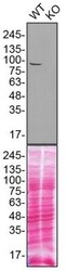
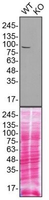
- Experimental details
- Western blot analysis of Dynamin 1 was performed by loading 130 µg of WT (lane 1) and DNM1 CRISPR KO (lane 2) U2OS cell lysates in RIPA buffer onto a 4-15% gradient polyacrylamide gel. Proteins were transferred to nitrocellulose membrane and blocked in 5% milk. Ponceau stained transfer of blot is shown. Dynamin 1 was detected at approximately 90 kDa using an Dynamin 1 monoclonal antibody (Product # MA5-15285) at a dilution of 1:1,000 in 5% BSA in TBST overnight at 4 deg, followed by secondary antibody diluted to 0.2 µg/mL using Goat anti-Mouse IgG (H+L) HRP antibody (Product # 62-6520). Chemiluminescent detection was performed using Pierce ECL Western Blotting Substrate (Product # 32106). Data courtesy of YCharOS Inc., an open science company with the mission of characterizing commercially available antibodies using knockout validation.
- Submitted by
- Invitrogen Antibodies (provider)
- Main image

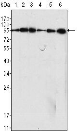
- Experimental details
- Western blot analysis of Dynamin-1 using Dynamin-1 monoclonal antibody (Product # MA5-15285) in C6 (1), NIH/3T3 (2), SKN-SH (3), LN18 (4), SHSY5Y (5) cell lysate and rat brain tissue lysate (6).
- Submitted by
- Invitrogen Antibodies (provider)
- Main image

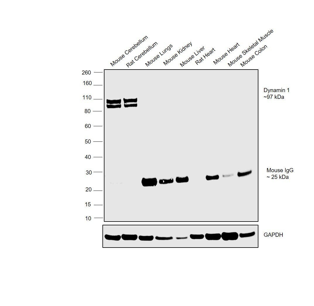
- Experimental details
- Western blot was performed using Anti-Dynamin 1 Monoclonal Antibody (3G4B6)(Product # MA5-15285) and a doublet band around 97 kDa band corresponding to Dynamin 1 was observed in Mouse and Rat Cerebellum tissues. Whole cell extracts (30 µg lysate) of Mouse Cerebellum (Lane 1), Rat Cerebellum (Lane 2), Mouse Lungs (Lane 3), Mouse Kidney (Lane 4), Mouse Liver (Lane 5), Rat Heart (Lane 6), Mouse Heart (Lane 7), Mouse Skeletal Muscle (Lane 8) and Mouse Colon (Lane 9) were electrophoresed using NuPAGE™ 4-12% Bis-Tris Protein Gel (Product # NP0321BOX). Resolved proteins were then transferred onto a Nitrocellulose membrane (Product # LC2001) by iBlot® 2 Dry Blotting System (Product # IB21001). The blot was probed with the primary antibody (1:1000 dilution) and detected by chemiluminescence with Goat anti-Mouse IgG (H+L) Superclonal™ Recombinant Secondary Antibody, HRP (Product # A28177, 1:4000 dilution) using the iBright FL 1000 (Product # A32752). Chemiluminescent detection was performed using Novex® ECL Chemiluminescent Substrate Reagent Kit (Product # WP20005).
Supportive validation
- Submitted by
- Invitrogen Antibodies (provider)
- Main image
- Experimental details
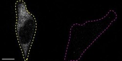
- Immunofluorescence of Dynamin-1 was performed using U2OS wild-type and DNM1 KO cells that were transfected with a green or a far red fluorescent dye, respectively. Post-transfection, WT and KO cells were mixed and plated to a 1:1 ratio on coverslips as a mosaic and incubated for 24 hrs. Cells were fixed in 4% PFA (in PBS) for 15 min and permeabilized with 0.1% Triton X-100. Cells were stained with the Dynamin-1 monoclonal antibody at a 1:1000 dilution overnight at 4 deg. Secondary antibody incubation was performed using 1 µg/mL of Goat anti-Mouse IgG (H+L) Highly Cross-Adsorbed Secondary Antibody, Alexa Fluor 555 antibody (Product # A21424) for 1 hr at RT. Imaging was performed with a 40X oil objective and analysis was performed using Image J. Cell image represents a single focal plane; WT and KO cells are outlined with a yellow (WT) or magenta (KO) dashed line. Data courtesy of YCharOS Inc., an open science company with the mission of characterizing commercially available antibodies using knockout validation.

Supportive validation
- Submitted by
- Invitrogen Antibodies (provider)
- Main image
- Experimental details
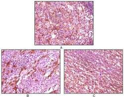
- Immunohistochemical analysis of paraffin-embedded human lymph tissue (A), glioma tissue (B) and cerebellum tissue (C) using Dynamin-1 monoclonal antibody (Product # MA5-15285) followed with DAB staining

Supportive validation
- Submitted by
- Invitrogen Antibodies (provider)
- Main image
- Experimental details
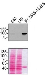
- Immunoprecipitation of Dynamin 1 was performed on U2OS cell lysates. Antibody-bead conjugates were prepared by adding 1 µg of Dynamin 1 monoclonal antibody (Product # MA5-15285) with 30 µL of protein G-Sepharose beads and rocked overnight at 4°C. 1 µg of DNM1 KO lysate was incubated with antibody-bead conjugate for 2 hrs at 4°C. After multiple washes, 10% starting material (SM), 10% unbound fraction (UB) and immunoprecipitated fraction (IP) were processed for immunoblot using a Dynamin 1 monoclonal antibody. Ponceau stained transfer of blot is shown. Data courtesy of YCharOS Inc., an open science company with the mission of characterizing commercially available antibodies using knockout validation.
