Antibody data
- Antibody Data
- Antigen structure
- References [2]
- Comments [0]
- Validations
- Western blot [1]
- Immunocytochemistry [1]
- Immunohistochemistry [1]
- Other assay [1]
Submit
Validation data
Reference
Comment
Report error
- Product number
- PA5-47075 - Provider product page

- Provider
- Invitrogen Antibodies
- Product name
- CD10 Polyclonal Antibody
- Antibody type
- Polyclonal
- Antigen
- Recombinant full-length protein
- Description
- In sandwich ELISAs, less than 35% cross-reactivity with recombinant human (rh) Neprilysin is observed and less than 0.2% cross-reactivity with rhNeprilysin-2 is observed. Reconstitute at 0.2 mg/mL in sterile PBS.
- Reactivity
- Human, Mouse, Rat
- Host
- Goat
- Isotype
- IgG
- Vial size
- 100 µg
- Concentration
- 0.2 mg/mL
- Storage
- -20° C, Avoid Freeze/Thaw Cycles
Submitted references HIF-1α and HIF-2α differently regulate tumour development and inflammation of clear cell renal cell carcinoma in mice.
Oncogenic HrasG12V expression plus knockdown of Cdkn2a using ecotropic lentiviral vectors induces high-grade endometrial stromal sarcoma.
Hoefflin R, Harlander S, Schäfer S, Metzger P, Kuo F, Schönenberger D, Adlesic M, Peighambari A, Seidel P, Chen CY, Consenza-Contreras M, Jud A, Lahrmann B, Grabe N, Heide D, Uhl FM, Chan TA, Duyster J, Zeiser R, Schell C, Heikenwalder M, Schilling O, Hakimi AA, Boerries M, Frew IJ
Nature communications 2020 Aug 17;11(1):4111
Nature communications 2020 Aug 17;11(1):4111
Oncogenic HrasG12V expression plus knockdown of Cdkn2a using ecotropic lentiviral vectors induces high-grade endometrial stromal sarcoma.
Brandt LP, Albers J, Hejhal T, Catalano A, Wild PJ, Frew IJ
PloS one 2017;12(10):e0186102
PloS one 2017;12(10):e0186102
No comments: Submit comment
Supportive validation
- Submitted by
- Invitrogen Antibodies (provider)
- Main image

- Experimental details
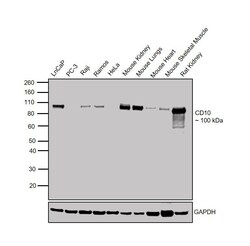
- Western blot was performed using Anti-CD10 Goat Polyclonal Antibody (Product # PA5-47075) and a 100 kDa band corresponding to CD10 was observed in cell lines and tissues tested except for PC-3 and HeLa. Membrane enriched extracts (30 µg lysate) of LNCaP (Lane 1), PC-3 (Lane 2), Raji (Lane 3), Ramos (Lane 4), HeLa (Lane 5), Mouse Kidney (Lane 6), Mouse Lungs (Lane 7), Mouse Heart (Lane 8), Mouse Skeletal Muscle (Lane 9) and Rat Kidney (Lane 10) were electrophoresed using Novex® NuPAGE® 4-12% Bis-Tris Protein Gel (Product # NP0322BOX). Resolved proteins were then transferred onto a nitrocellulose membrane (Product # IB23001) by iBlot® 2 Dry Blotting System (Product # IB21001). The blot was probed with the primary antibody (0.1 µg/mL) and detected by chemiluminescence with Rabbit anti-Goat IgG (H+L) Superclonal™ Recombinant Secondary Antibody, HRP (Product # A27014, 1:4000 dilution) using the iBright FL 1000 (Product # A32752). Chemiluminescent detection was performed using Novex® ECL Chemiluminescent Substrate Reagent Kit (Product # WP20005).
Supportive validation
- Submitted by
- Invitrogen Antibodies (provider)
- Main image

- Experimental details
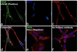
- Immunofluorescence analysis of CD10 was performed using LNCaP and HeLa cells. The cells were fixed with 4% paraformaldehyde for 10 minutes and blocked with 2% BSA for 1 hour at room temperature. The cells were labeled with CD10 Goat Polyclonal Antibody (Product # PA5-47075) at 5 µg/mL in 0.1% BSA and incubated overnight at 4 degree and then labeled with Rabbit anti-Goat IgG (H+L) Superclonal™ Recombinant Secondary Antibody, Alexa Fluor® 488 (Product # A27012) at a dilution of 1:2000 for 45 minutes at room temperature (Panel a: green) in LNCaP cells. Nuclei (Panel b: blue) were stained with ProLong™ Diamond Antifade Mountant with DAPI (Product # P36962). F-actin (Panel c: red) was stained with Rhodamine Phalloidin (Product # R415, 1:300). Panel d represents the merged image of LNCaP cells, which is a positive model for CD10 expression showing a plasma membrane localization. Panel e represents the merged image of HeLa cells, that are null for CD10 protein expression. Panel f represents control cells with no primary antibody to assess background. The images were captured at 60X magnification.
Supportive validation
- Submitted by
- Invitrogen Antibodies (provider)
- Main image

- Experimental details
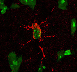
- Immunohistochemical analysis of CD10 in perfusion fixed frozen sections of mouse brain (glial cell in hippocampus). Samples were incubated in CD10 polyclonal antibody (Product # PA5-47075) using a dilution of 15 µg/mL overnight at 4 °C. Tissue was stained (red) and counterstained (green).
Supportive validation
- Submitted by
- Invitrogen Antibodies (provider)
- Main image

- Experimental details
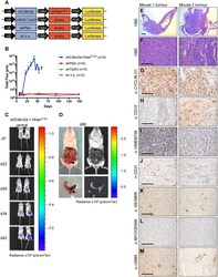
- Fig 4 Hras G12V expression plus knockdown of Cdkn2a causes high-grade endometrial stromal sarcomas. ( A ) Schematic of MuLE vectors simultaneously expressing a combination of shRNA against Cdkn2a plus expression of Hras G12V or expressing shRNAs against Trp53 or Pten or a non-silencing (n.s.) control shRNA. All vectors also expressed Luciferase. Numbers of mice that were injected with each vector are shown in the figure. ( B ) Quantification (mean +- SD) of luciferase signal over time. + Sacrifice of all mice in this group by this time point. ( C ) Bioluminescence imaging in two mice 7, 22, 28, 34 and 42 days after the injection of MuLE lentiviruses expressing shRNA against Cdkn2a together with Hras G12V into the uterus of 6-8-week-old SCID/beige mice. Injected mice developed tumours (n = 4) with 80% penetrance. Median overall survival was 49 days. ( D ) Bioluminescence imaging and photographs of tumour-bearing uterus at the time of sacrifice (day 56 after injection). ( E - M ) H&E and immunohistochemical stainings of tumours from two different mice using the indicated antibodies. Low magnification scale bar: 1 mm and high magnification scale bar: 100 mum.
 Explore
 Validate
 Learn
Learn Western blot
 ELISA
ELISA