Antibody data
- Antibody Data
- Antigen structure
- References [2]
- Comments [0]
- Validations
- Western blot [2]
- Immunohistochemistry [2]
Submit
Validation data
Reference
Comment
Report error
- Product number
- PA1-28838 - Provider product page

- Provider
- Invitrogen Antibodies
- Product name
- GLI2 Polyclonal Antibody
- Antibody type
- Polyclonal
- Antigen
- Synthetic peptide
- Description
- PA1-28838 detects Gli2 from mouse samples.
- Reactivity
- Human, Mouse
- Host
- Rabbit
- Isotype
- IgG
- Vial size
- 50 µg
- Concentration
- 1.02 mg/mL
- Storage
- Store at 4°C short term. For long term storage, store at -20°C, avoiding freeze/thaw cycles.
Submitted references Suppressor of fused controls cerebellum granule cell proliferation by suppressing Fgf8 and spatially regulating Gli proteins.
Novel mTORC1 Mechanism Suggests Therapeutic Targets for COMPopathies.
Jiwani T, Kim JJ, Rosenblum ND
Development (Cambridge, England) 2020 Feb 3;147(3)
Development (Cambridge, England) 2020 Feb 3;147(3)
Novel mTORC1 Mechanism Suggests Therapeutic Targets for COMPopathies.
Posey KL, Coustry F, Veerisetty AC, Hossain MG, Gambello MJ, Hecht JT
The American journal of pathology 2019 Jan;189(1):132-146
The American journal of pathology 2019 Jan;189(1):132-146
No comments: Submit comment
Supportive validation
- Submitted by
- Invitrogen Antibodies (provider)
- Main image

- Experimental details
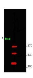
- Western blot analysis of Gli2 in mouse brain whole cell lysate using a Gli2 polyclonal antibody (Product # PA1-28838) at a dilution of 1:750. Results show detection of a predominant band at ~190 kDa corresponding to Gli-2 (arrowhead) in lane 1. Molecular weight markers are shown (M) using the 700 nm channel (red).
- Submitted by
- Invitrogen Antibodies (provider)
- Main image

- Experimental details
- Western blot using GLI2 Polyclonal Antibody (Product # PA1-28838) shows detection of a predominant band at 190 kDa corresponding to Gli-2 (arrowhead) in mouse brain whole cell lysate (lane 1). Pre-incubation of antibody with immunizing peptide completely blocks staining of this band (lane 2). 25 µg of lysate was resolved on a 4-8% Tris-glycine gel by SDS-PAGE and transferred onto nitrocellulose. After blocking with 5% goat serum and 0.5% BLOTTO in PBS, the membrane was probed with the primary antibody diluted to 1:750. Incubation was at room temperature for 2 h followed by washes and reaction with a 1:10,000 dilution of a Gt-a-Rabbit IgG (H&L) MX10 for 45 min at room temperature. Molecular weight markers are shown (M) using the 700 nm channel (red).
Supportive validation
- Submitted by
- Invitrogen Antibodies (provider)
- Main image

- Experimental details
- Immunohistochemistry analysis of GLI2 was performed in several formalin-fixed and paraffin embedded mouse brain using GLI2 Polyclonal Antibody (Product # PA1-28838) at 10 µg/mL. Moderate to strong staining was seen on many tissues with low background staining. This image shows Gli-2 staining of mouse brain.
- Submitted by
- Invitrogen Antibodies (provider)
- Main image

- Experimental details
- Immunohistochemistry analysis of GLI2 was performed in several formalin-fixed and paraffin embedded mouse testis using GLI2 Polyclonal Antibody (Product # PA1-28838) at 10 µg/mL. Moderate to strong staining was seen on many tissues with low background staining. This image shows Gli-2 staining of mouse testis.
 Explore
 Validate
 Learn
Learn Western blot
 ELISA
ELISA