Explore
 Validate
 Learn
LearnMA5-25317
antibody from Invitrogen Antibodies
Targeting: HDAC6
FLJ16239, HD6, JM21, KIAA0901, PPP1R90
 Western blot
 Flow cytometry
Antibody data
- Antibody Data
- Antigen structure
- References [0]
- Comments [0]
- Validations
- Western blot [3]
- Immunocytochemistry [4]
- Immunohistochemistry [4]
- Chromatin Immunoprecipitation [1]
Submit
Validation data
Reference
Comment
Report error
- Product number
- MA5-25317 - Provider product page

- Provider
- Invitrogen Antibodies
- Product name
- HDAC6 Monoclonal Antibody (OTI4C5)
- Antibody type
- Monoclonal
- Antigen
- Recombinant full-length protein
- Reactivity
- Human, Rat, Canine
- Host
- Mouse
- Isotype
- IgG
- Antibody clone number
- OTI4C5
- Vial size
- 100 µL
- Concentration
- 1 mg/mL
- Storage
- -20° C, Avoid Freeze/Thaw Cycles
No comments: Submit comment
Supportive validation
- Submitted by
- Invitrogen Antibodies (provider)
- Main image
- Experimental details
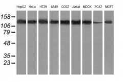
- Western blot analysis of HDAC6 in HepG2, HeLa, HT29, A549, COS7, Jurkat, MDCK, PC12, MCF7 cells using 35 µg per lane. Samples were probed with HDAC6 (Product # MA5-25317) monoclonal antibody.

- Submitted by
- Invitrogen Antibodies (provider)
- Main image
- Experimental details
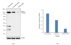
- Knockdown of HDAC6 was achieved by transfecting A549 with HDAC6 specific siRNAs (Silencer® select Product # s19459). Western blot analysis (Fig. a) was performed using whole cell extracts from the HDAC6 knockdown cells (lane 3), non-specific scrambled siRNA transfected cells (lane 2) and untransfected cells (lane 1). The blot was probed with HDAC6 Monoclonal Antibody (OTI4C5) (Product # MA5-25317, 1:1000 dilution) and Goat anti-Mouse IgG (H+L), Superclonal™ Recombinant Secondary Antibody, HRP conjugate (Product # A28177, 1:4000 dilution). Densitometric analysis of this western blot is shown in histogram (Fig. b). Decrease in signal upon siRNA mediated knock down confirms that antibody is specific to HDAC6.

- Submitted by
- Invitrogen Antibodies (provider)
- Main image
- Experimental details
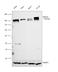
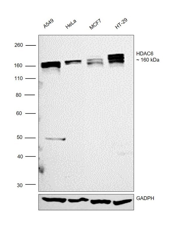
- Western blot was performed using Anti-HDAC6 Monoclonal Antibody (OTI4C5) (Product # MA5-25317) and a 160kDa band corresponding to HDAC6 was observed in the cell lines tested. Whole cell extracts of A549 (Lane 1), HeLa (Lane 2), MCF7 (Lane 3) and HT-29 (Lane 4) were electrophoresed using Novex® NuPAGE® 4-12 % Bis-Tris gel (Product # NP0321BOX). Resolved proteins were then transferred onto a nitrocellulose membrane (Product # IB23001) by iBlot® 2 Dry Blotting System (Product # IB21001). The blot was probed with the primary antibody (1:1000 dilution) and detected by Goat anti-Mouse IgG (H+L), Superclonal™ Recombinant Secondary Antibody, HRP conjugate (Product # A28177, 1:4000 dilution) using the iBright FL 1000 (Product # A32752). Chemiluminescent detection was performed using Novex® ECL Chemiluminescent Substrate Reagent Kit (Product # WP20005).
Supportive validation
- Submitted by
- Invitrogen Antibodies (provider)
- Main image

- Experimental details
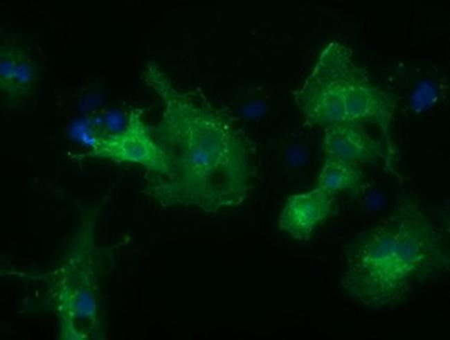
- Immunofluorescent analysis of HDAC6 in COS7 cells. Cells were transfected with a plasmid overexpressing HDAC6 and probed with a HDAC6 monoclonal antibody (Product # MA5-25317).
- Submitted by
- Invitrogen Antibodies (provider)
- Main image

- Experimental details
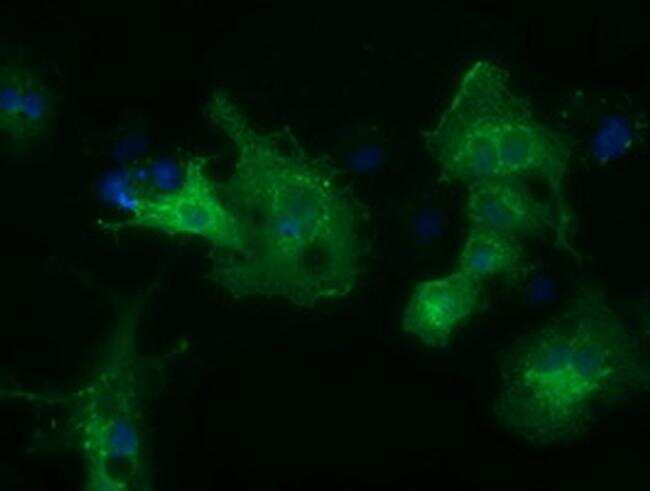
- Immunofluorescent analysis of HDAC6 in COS7 cells. Cells were transfected with a plasmid overexpressing HDAC6 and probed with a HDAC6 monoclonal antibody (Product # MA5-25317).
- Submitted by
- Invitrogen Antibodies (provider)
- Main image

- Experimental details
- Immunofluorescent analysis of HDAC6 in COS7 cells. Cells were transfected with a plasmid overexpressing HDAC6 and probed with a HDAC6 monoclonal antibody (Product # MA5-25317).

- Submitted by
- Invitrogen Antibodies (provider)
- Main image
- Experimental details
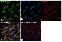
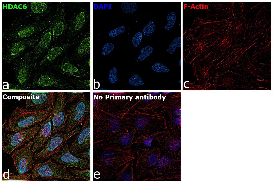
- Immunofluorescence analysis of HDAC6 was performed using 70% confluent log phase HeLa cells. The cells were fixed with 4% paraformaldehyde for 10 minutes, permeabilized with 0.1% Triton™ X-100 for 15 minutes, and blocked with 2% BSA for 1 hour at room temperature. The cells were labeled with HDAC6 Mouse Monoclonal Antibody (Product # MA5-23517) at 1:100 dilution in 0.1% BSA and incubated overnight at 4 degree and then labeled with Goat anti-Mouse IgG (H+L) Superclonal™ Recombinant Secondary Antibody, Alexa Fluor® 488 conjugate (Product # A28175) at a dilution of 1:2000 for 45 minutes at room temperature (Panel a: green). Nuclei (Panel b: blue) were stained with ProLong™ Diamond Antifade Mountant with DAPI (Product # P36962). F-actin (Panel c: red) was stained with Rhodamine Phalloidin (Product # R415, 1:300). Panel d represents the composite image showing nuclear and cytoplasmic localization of HDAC6. Panel e represents control cells with no primary antibody to assess background. The images were captured at 60X magnification.
Supportive validation
- Submitted by
- Invitrogen Antibodies (provider)
- Main image

- Experimental details
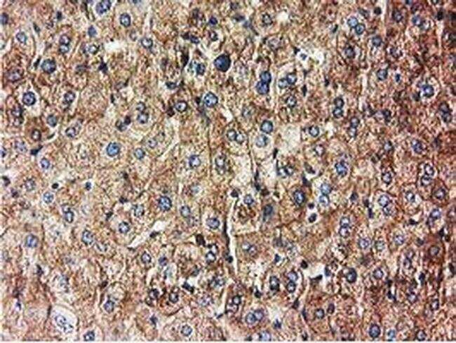
- Immunohistochemical analysis of HDAC6 in paraffin-embedded human liver tissue using a HDAC6 monoclonal antibody (Product # MA5-25317) at a dilution of 1:150.
- Submitted by
- Invitrogen Antibodies (provider)
- Main image

- Experimental details
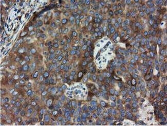
- Immunohistochemical analysis of HDAC6 in paraffin-embedded adenocarcinoma of human ovary tissue using a HDAC6 monoclonal antibody (Product # MA5-25317) at a dilution of 1:150.
- Submitted by
- Invitrogen Antibodies (provider)
- Main image
- Experimental details
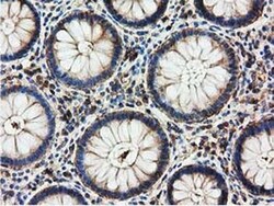
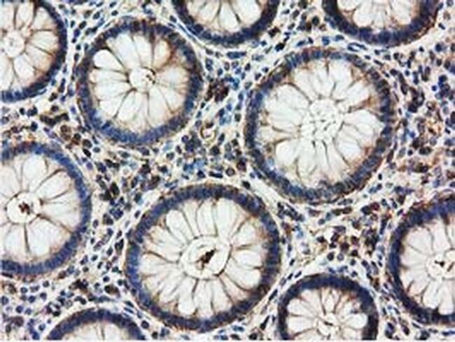
- Immunohistochemical analysis of HDAC6 in paraffin-embedded human colon tissue using a HDAC6 monoclonal antibody (Product # MA5-25317) at a dilution of 1:150.
- Submitted by
- Invitrogen Antibodies (provider)
- Main image

- Experimental details
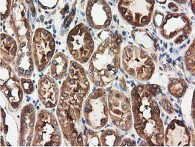
- Immunohistochemical analysis of HDAC6 in paraffin-embedded human kidney tissue using a HDAC6 monoclonal antibody (Product # MA5-25317) at a dilution of 1:150.
Supportive validation
- Submitted by
- Invitrogen Antibodies (provider)
- Main image

- Experimental details
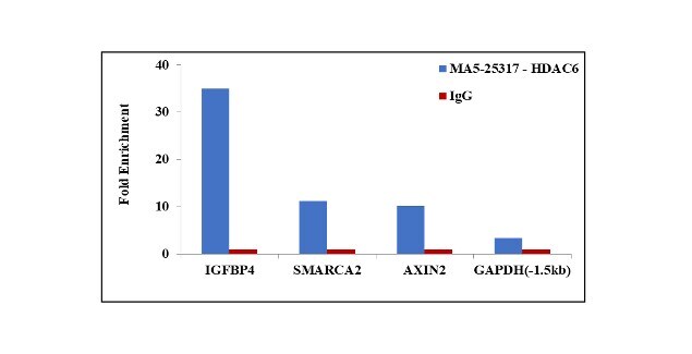
- Chromatin Immunoprecipitation (ChIP) assay of endogenous HDAC6 protein using Anti-HDAC6 Antibody: ChIP was performed using Anti-HDAC6 Mouse Monoclonal Antibody (Product # MA5-25317, 5 µg) on sheared chromatin from HeLa cells using the MAGnify ChIP System kit (Product # 49-2024). Normal Mouse IgG was used as a negative IP control. The purified DNA was analyzed by qPCR using primers binding to IGFBP4 promoter (-4.2kb), SMARCA2 transcriptional start site, AXIN2 exon1 and GAPDH promoter. Data is presented as fold enrichment of the antibody signal versus the negative control IgG using the comparative CT method.