Explore
 Validate
 Learn
LearnPA5-84083
antibody from Invitrogen Antibodies
Targeting: CAMK2A
CAMKA, CaMKIINalpha, CaMKIIα, KIAA0968
 Western blot
Antibody data
- Antibody Data
- Antigen structure
- References [1]
- Comments [0]
- Validations
- Western blot [2]
- Immunocytochemistry [1]
- Immunohistochemistry [1]
- Other assay [1]
Submit
Validation data
Reference
Comment
Report error
- Product number
- PA5-84083 - Provider product page

- Provider
- Invitrogen Antibodies
- Product name
- CaMKII alpha Polyclonal Antibody
- Antibody type
- Polyclonal
- Antigen
- Recombinant full-length protein
- Reactivity
- Human
- Host
- Rabbit
- Isotype
- IgG
- Vial size
- 100 µL
- Concentration
- 0.1 mg/mL
- Storage
- Store at 4°C short term. For long term storage, store at -20°C, avoiding freeze/thaw cycles.
Submitted references Pervasive compartment-specific regulation of gene expression during homeostatic synaptic scaling.
Colameo D, Rajman M, Soutschek M, Bicker S, von Ziegler L, Bohacek J, Winterer J, Germain PL, Dieterich C, Schratt G
EMBO reports 2021 Oct 5;22(10):e52094
EMBO reports 2021 Oct 5;22(10):e52094
No comments: Submit comment
Supportive validation
- Submitted by
- Invitrogen Antibodies (provider)
- Main image
- Experimental details
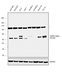
- Western blot was performed using Anti-CaMKII alpha Polyclonal Antibody, (Product # PA5-84083) and a ~54 kDa band corresponding to CaMKII alpha was observed across the panel tested except SK-BR-3 and OVCAR-3 which are reported to be negative. An uncharacterized band (*) at ~100kDa was also observed across the cell lines tested. Whole cell extracts (30 µg lysate) of U-87 MG (Lane 1), SK-N-SH (Lane 2), PC-3 (Lane 3), THP-1 (Lane 4), SK-BR-3 (Lane 5), OVCAR-3 (Lane 6), Neuro-2a (Lane 7) and PC-12 (Lane 8) were electrophoresed using Novex® NuPAGE® 4-12% % Bis-Tris gel (Product # NP0322BOX). Resolved proteins were then transferred onto a nitrocellulose membrane (Product # IB23001) by iBlot® 2 Dry Blotting System (Product # IB21001). The blot was probed with the primary antibody (1:500 dilution) and detected by chemiluminescence with Goat anti-Rabbit IgG (H+L) Superclonal™ Recombinant Secondary Antibody, HRP (Product # A27036, 1:4000 dilution) using the iBright FL 1000 (Product # A32752). Chemiluminescent detection was performed using Novex® ECL Chemiluminescent Substrate Reagent Kit (Product # WP20005).
- Submitted by
- Invitrogen Antibodies (provider)
- Main image
- Experimental details
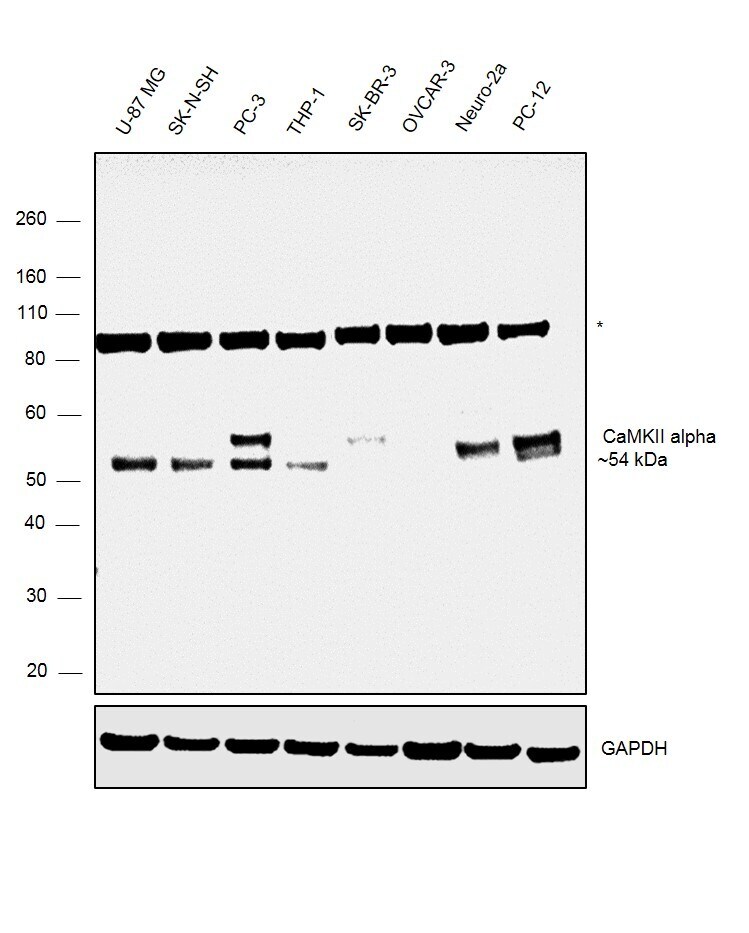
- Western blot was performed using Anti-CaMKII alpha Polyclonal antibody, (Product # PA5-84083) and a ~54 kDa band corresponding to CaMKII alpha was observed in Mouse and Rat Brain but not in Mouse, Rat Lung and Heart which are reported to be negative. Two uncharacterized bands (*), one at ~60kDa was observed in all the tissues and another at ~80kDa was observed in all the mouse tissues tested. Tissue extracts (30 µg lysate) of Mouse Brain (Lane1), Mouse Lung (Lane 2), Mouse Heart (Lane 3), Rat Brain (Lane 4), Rat Lung (Lane 5) and Rat Heart (Lane 6) were electrophoresed using Novex® NuPAGE® 4-12 % Bis-Tris gel (Product # NP0322BOX). Resolved proteins were then transferred onto a nitrocellulose membrane (Product # IB23001) by iBlot® 2 Dry Blotting System (Product # IB21001). The blot was probed with the primary antibody (1:500 dilution) and detected by chemiluminescence with Goat anti-Rabbit IgG (H+L) Superclonal™ Recombinant Secondary Antibody, HRP (Product # A27036, 1:4000 dilution) using the iBright FL 1000 (Product # A32752). Chemiluminescent detection was performed using Novex® ECL Chemiluminescent Substrate Reagent Kit (Product # WP20005).

Supportive validation
- Submitted by
- Invitrogen Antibodies (provider)
- Main image
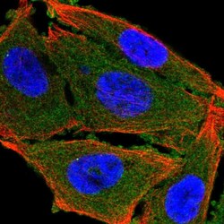
- Experimental details
- Immunofluorescent analysis of CaMKII alpha in PC-3 cells using a CaMKII alpha polyclonal antibody (Product # PA5-84083). The analysis shows localization to plasma membrane & cytosol.
Supportive validation
- Submitted by
- Invitrogen Antibodies (provider)
- Main image
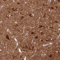
- Experimental details
- Immunohistochemical analysis of CaMKII alpha in human cerebral cortex using a CaMKII alpha polyclonal antibody (Product # PA5-84083). The analysis shows strong cytoplasmic positivity in neuronal cells.
Supportive validation
- Submitted by
- Invitrogen Antibodies (provider)
- Main image
- Experimental details
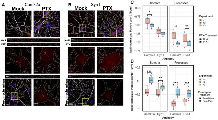
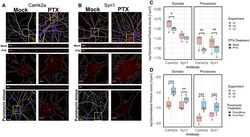
- Figure 5 PTX-mediated regulation of local translation Representative pictures of nascent Camk2a-peptides in mock- and PTX-treated neurons. Upper Panels: Representative overview images with merged channels (grey: MAP2-staining, red: Duolink-PLA signal, blue: Hoechst nuclear staining). Middle Panels: Magnifications of boxed areas marked in (A). MAP2 signal was used to outline somata and dendritic processes (white lines). Lower Panels: Negative control of puromycin-untreated cells with representative Duolink signal in dendrite and soma, respectively. (Scale bars: in overview images 20 mum, in inserts of higher magnification 5 mum). Same as (A) with nascent Synapsin1-peptides. Quantification of Duolink-particle density of (A) and (B) assessing PTX treatment effect. n = 3 independent experiments with 10 images averaged per condition and experiment; Three-way ANOVA followed by Tukey's post hoc multiple comparison test; * P < 0.05; ** P < 0.01). Quantification of Duolink-particle density of (A) and (B) assessing Puromycin-treatment effect. n = 3 independent experiments for Puromycin-Minus and n = 6 data points for Puromycin-Plus conditions (PTX and mock conditions combined together across n = 3 independent experiments, taken into account in ANOVA model) with 10 images averaged per condition and experiment; three-way ANOVA followed by Tukey's post hoc multiple comparison test; ** P < 0.01, *** P < 0.001). Data information: Boxplots: central line: median; box: 25 th to 75 th percentil